
TABLE OF CONTENTS
Bovine Theileriosis (Theileria annulata): Life Cycle, Diagnosis & Treatment
Theileria annulata is a tick-borne protozoan parasite that causes bovine theileriosis, a major hemoprotozoan disease of cattle and buffaloes. The disease is transmitted primarily by Hyalomma ticks and is characterized by fever, enlargement of superficial lymph nodes, anemia, reduced milk production, and high mortality in susceptible animals. Endemic in many tropical and subtropical regions, bovine theileriosis results in significant economic losses due to decreased productivity and increased treatment costs.
Parasite Overview
- Host: Cattle and buffaloes. However, buffaloes usually act as reservoir hosts.
- Vector: Hyalomma anatolicum anatolicum and H. marginatum isaaci
- Transmission: Transtadial transmission (stage-to-stage)
Morphology and Predilection Site
Piroplasms or Merozoites
Piroplasms are found in erythrocytes. They are pleomorphic; round, oval, ring (signet ring), rod, comma, and Anaplasma-like forms are seen. When stained with Romanowsky stain, they appear as blue cytoplasm with red chromatin granules at one end.
The merozoites in the erythrocytes multiply by binary fission to produce two daughter cells, which in turn divide to form four cells. Hence, multiple infections of RBCs with theilerial organisms are common.
Schizonts (Koch’s Blue Bodies – KBB)
The schizont of Theileria species is called Koch’s blue body, which is the actively multiplying and most pathogenic stage of the parasite. Koch’s blue bodies are found in the lymphocytes of the spleen and lymph nodes, and in severe infections, they may also be found free in the blood.
They are circular or irregularly shaped, measuring approximately 8 µm in diameter and up to 15–27 µm. During the initial stage of multiplication, the schizonts are large and are called macroschizonts. However, following further nuclear division, they become smaller and are called microschizonts.
The microschizonts, in turn, produce merozoites. These merozoites invade RBCs and form intraerythrocytic merozoites (piroplasms), which later develop into spherical or ovoid gamonts. The gamonts are transformed into gametes within the tick following a blood meal.
T. annulata is readily transmitted through blood because numerous schizonts are found in the circulating blood.
Life Cycle
Transtadial (stage-to-stage) transmission occurs. If larvae acquire the infection from diseased animals while feeding, the nymphal stage will be able to transmit the infection to susceptible hosts. Congenital infections rarely occur in calves.
Development of the Parasite in the Vertebrate Host
When an infected tick bites a susceptible host, sporozoites of T. annulata are inoculated with saliva after 2–4 days of attachment. The inoculated sporozoites enter lymphocytes located near the site of the tick bite and are then carried to adjacent lymph nodes, such as the prescapular, parotid, submandibular, and precrural lymph nodes.
In the lymph nodes, the sporozoites undergo multiplication by schizogony. These multiplying parasites stimulate lymphoproliferation, leading to rapid division of lymphocytes.
When an infected lymphocyte divides into two, the parasites also divide and infect both newly formed lymphocytes. This process is called clonal expansion and is responsible for enlargement of the lymph nodes, particularly the prescapular lymph node, in theileriosis.
The developmental stage found in the lymphocytes is called the schizont (meront) or KBB, which produces merozoites. Infected animals develop high fever during schizont formation and the release of merozoites from the lymphocytes.
The released merozoites enter erythrocytes, where they multiply by binary fission and eventually develop into spherical or ovoid gamonts.
Development of the Parasite in the Vector
When a larva or nymph feeds on infected animals, gamonts are ingested with the blood meal. Following lysis of RBCs in the tick’s gut, the gamonts are released and differentiate into macrogamonts and microgamonts, which subsequently produce macrogametes and microgametes, respectively.
The macrogametes and microgametes (Ray bodies) unite to form a zygote within 12–13 days after the blood meal. The zygotes then enter the cells of the gut wall, where they develop into elongated, club-shaped, motile kinetes (ookinetes).
These ookinetes then leave the gut wall and enter the hemolymph, from where they migrate to the salivary glands by days 17–20. By this time, the ticks molt to the next stage; however, further development of the parasite occurs only when the ticks attach to a new host, followed by activation of the salivary gland acinar cells.
In the activated acinar cells, the ookinetes undergo nuclear division to form a young sporont, followed by sporoblasts, which in turn divide to produce thousands of comma-shaped sporozoites.
These sporozoites are released into the saliva when the sporoblasts rupture and are inoculated into the host when the tick bites, usually 2 days after attachment.
Mode of Transmission (Stage-to-Stage Transmission)
Infected animals → Larva (infected) → Nymph (transmits) → New host
Infected animals → Nymph (infected) → Adult (transmits) → New host.
Epizootiology
The prevalence of theileriosis depends on the prevalence of tick vectors. Stress caused by hot and humid weather, transportation, intercurrent infections, and vaccination may predispose animals to theileriosis.
The incidence of theileriosis is high during the monsoon season. Native (indigenous) cattle mostly act as carriers and rarely show clinical signs, whereas exotic breeds such as Holstein Friesian and Jersey are more susceptible. Mortality may reach 70–80%, and morbidity may reach 90–100%.
Buffaloes are generally resistant and act as reservoir hosts. Newborn calves may also be susceptible to this disease. The disease may become enzootic once it occurs in a particular locality. It is very difficult to eradicate unless its life cycle is interrupted.
Pathogenesis and Clinical Signs
Clinical manifestations of theileriosis are classified into four forms.
1. Peracute Form
The animal dies within 3–4 days of infection following a period of high fever, although this form is uncommon.
2. Acute Form
This is the most common form of the disease and is characterized by high fever (104–107 °F) as the first sign, inappetence, cessation of rumination, tachycardia, weakness, lacrimation, decreased milk production, swelling of superficial lymph nodes (prescapular/precrural) and eyelids, diarrhea containing blood and mucus, icteric mucous membranes, and petechial hemorrhages.
Marked anemia is present, but hemoglobinuria is absent. Incoordination, teeth grinding, and urticarial skin lesions may also be observed in some cases. Affected animals become emaciated, and death may occur.
3. Subacute Form
There is an irregular intermittent fever that lasts for 10–15 days, after which animals may recover spontaneously. Abortion may occur in pregnant animals.
4. Chronic Form
There is intermittent fever, inappetence, marked emaciation, anemia, and icterus. In some cases, due to stress, the chronic form of the disease may progress to the acute form, and animals may die within 1–2 days.
Postmortem Lesions
- Splenomegaly
- Hepatomegaly with a distended gallbladder
- Renal infarction
- Pulmonary edema — the main cause of death in theileriosis
- Icteric mucous membranes with petechiae
- The abomasum and the small and large intestines are swollen and contain characteristic punched-out necrotic ulcers surrounded by a zone of inflammation.
Immunity
Premunity develops due to the presence of residual parasites in the host.
Diagnosis
- History of transportation, calving, and vaccination.
- Prevalence of tick vectors, season, and locality where theileriosis is endemic.
- Clinical signs.
- Demonstration of piroplasms in blood smears and schizonts (KBB) in lymph node smears.
Treatment
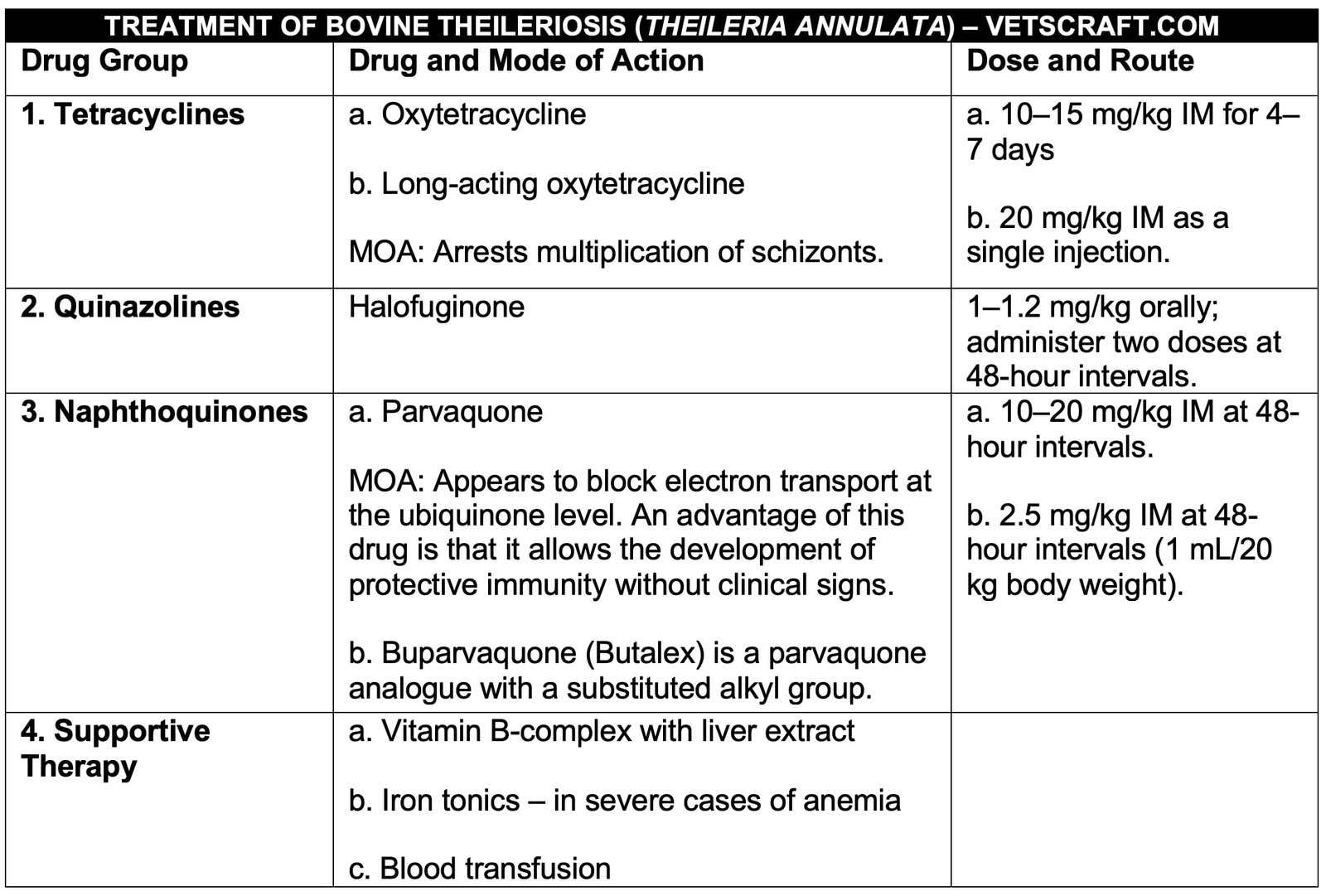
Control of Theileriosis
Tick Control
Use various classes of insecticides such as deltamethrin, flumethrin, and cypermethrin at a concentration of 0.1–0.2%.
Vaccination
Rakshavac-T vaccine, produced by Indian Immunologicals Ltd., Hyderabad, is used for immunization of cattle against theileriosis. It contains schizont-infected lymphocytes (SIL) attenuated through 150 passages at a concentration of 10⁶ cells/dose. The vaccine should be stored in liquid nitrogen (LN₂), and the reconstituted vaccine must be used within 2 hours.
- Package: 2 doses — Volume: 1 mL — Diluent: 5 mL — Dose: 3 mL
- Package: 5 doses — Volume: 2.5 mL — Diluent: 12.5 mL — Dose: 3 mL
Annual vaccination is recommended in endemic areas.
- Age of Vaccination: Adults and calves older than 3 months
- Route of Administration: Subcutaneous
- Duration of Immunity: 3 years
Administration
Remove the vaccine vial from the liquid nitrogen container and thaw the vaccine concentrate in lukewarm water (37°C). Transfer the thawed vaccine concentrate using a sterile needle and syringe into the corresponding vaccine diluent vial and mix gently. The reconstituted vaccine should be injected subcutaneously into the shoulder or dewlap.
Precautions
- Do not vaccinate animals in the advanced stage of pregnancy.
- The reconstituted vaccine should be used immediately and should not be frozen for reuse.

