
Vomer Bone
Veterinary AnatomyVomer bone of ox is a “U” or “V” shaped median bone, which forms the ventral part of the septum nasi. It is composed of a thin lamina.
Vomer bone of ox is a “U” or “V” shaped median bone, which forms the ventral part of the septum nasi. It is composed of a thin lamina.
Turbinate bone are delicate scroll like bones two on either side, attached to the lateral walls of the nasal cavity.
The malar bone or zygomatic bone is irregularly triangular and is situated between the lacrimal and maxilla
Lacrimal bone of Ox are situated at the anterior part of the orbits and extend forward on the face
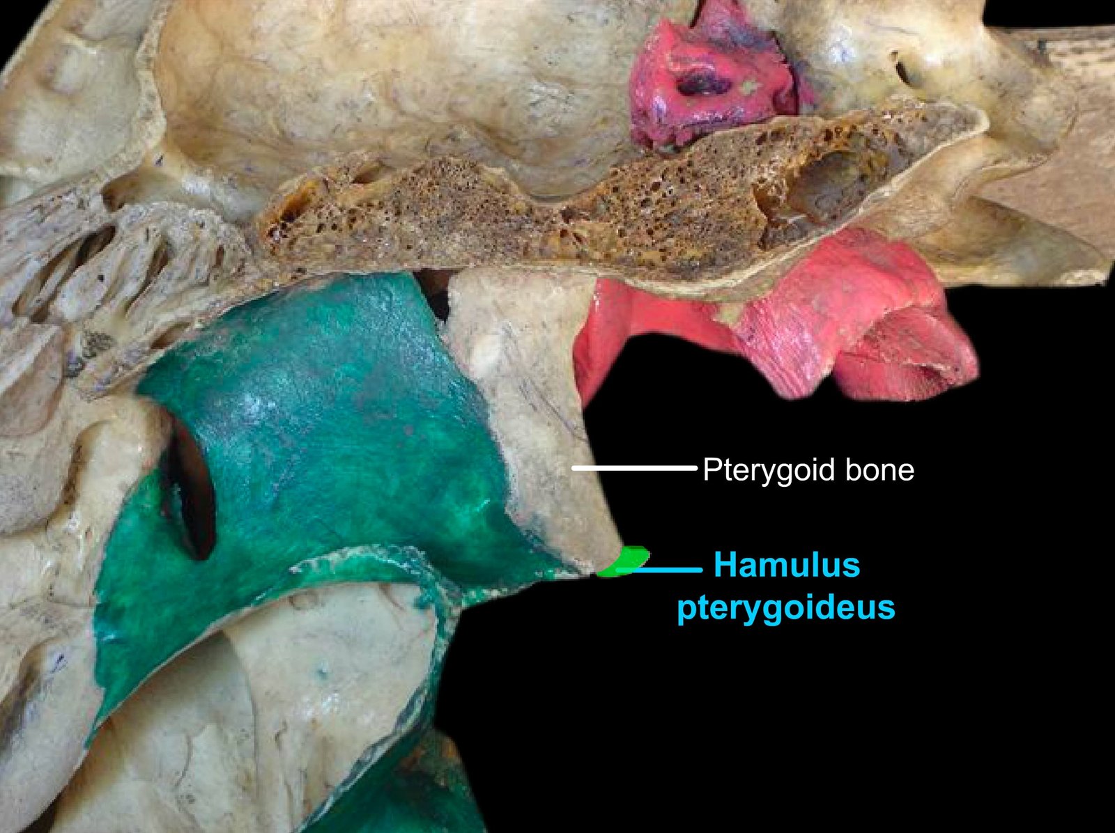
Learn the anatomy of the pterygoid bone in ox, horse, dog, and fowl. Study its location, articulations, pterygoid canal formation, and species differences in veterinary anatomy.
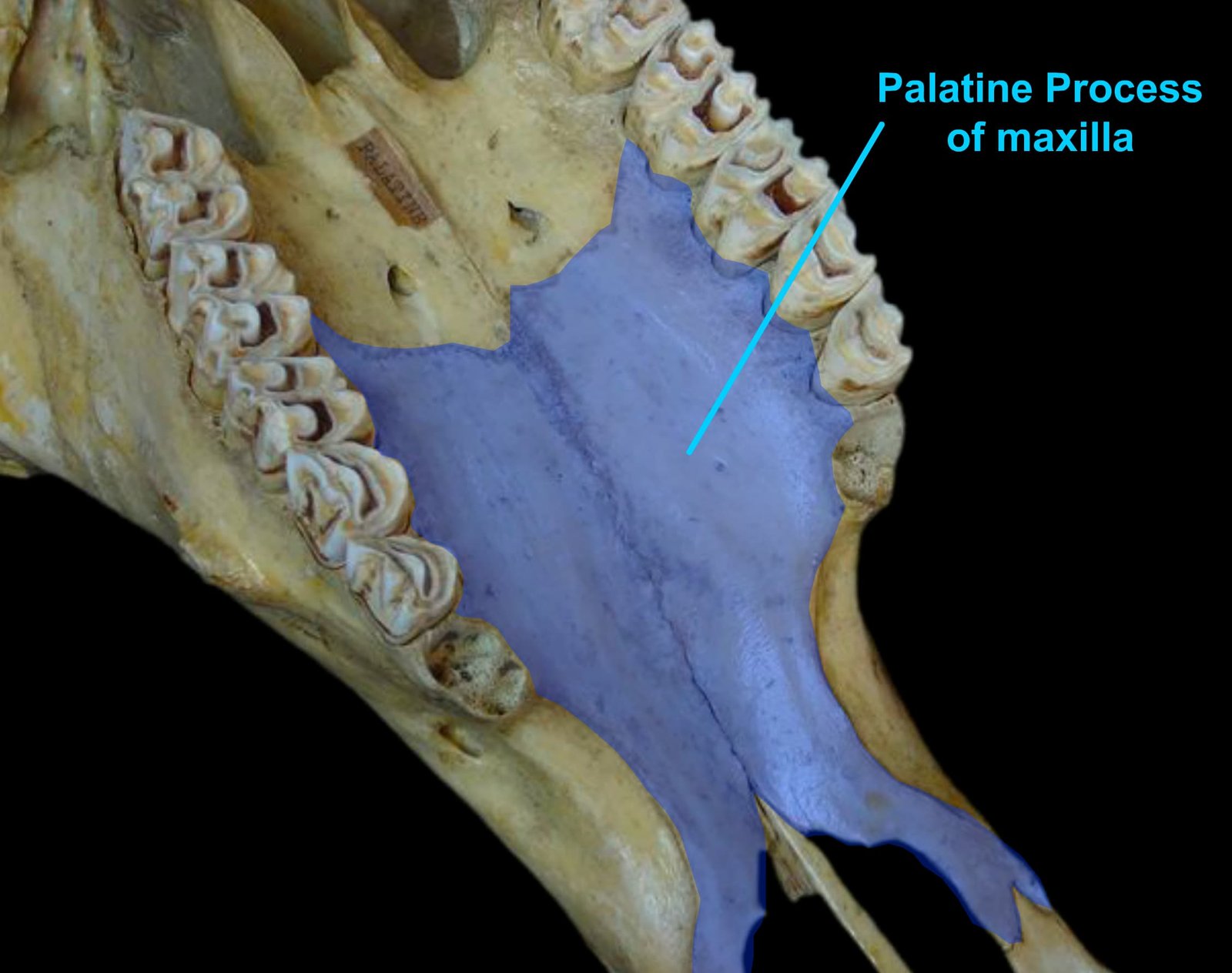
Learn the anatomy of the palatine bone in ox, horse, dog, and fowl, including horizontal and vertical parts, foramina, articulations, and species differences in veterinary anatomy.
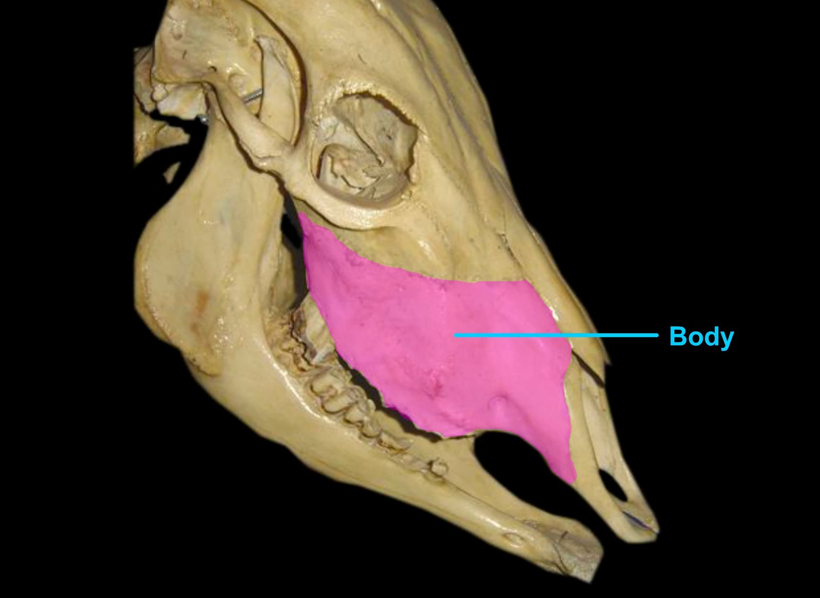
Learn the anatomy of the maxilla bone in ox, horse, dog, and fowl with comparative veterinary anatomy notes including facial crest, infraorbital foramen, palatine process, and important species differences.
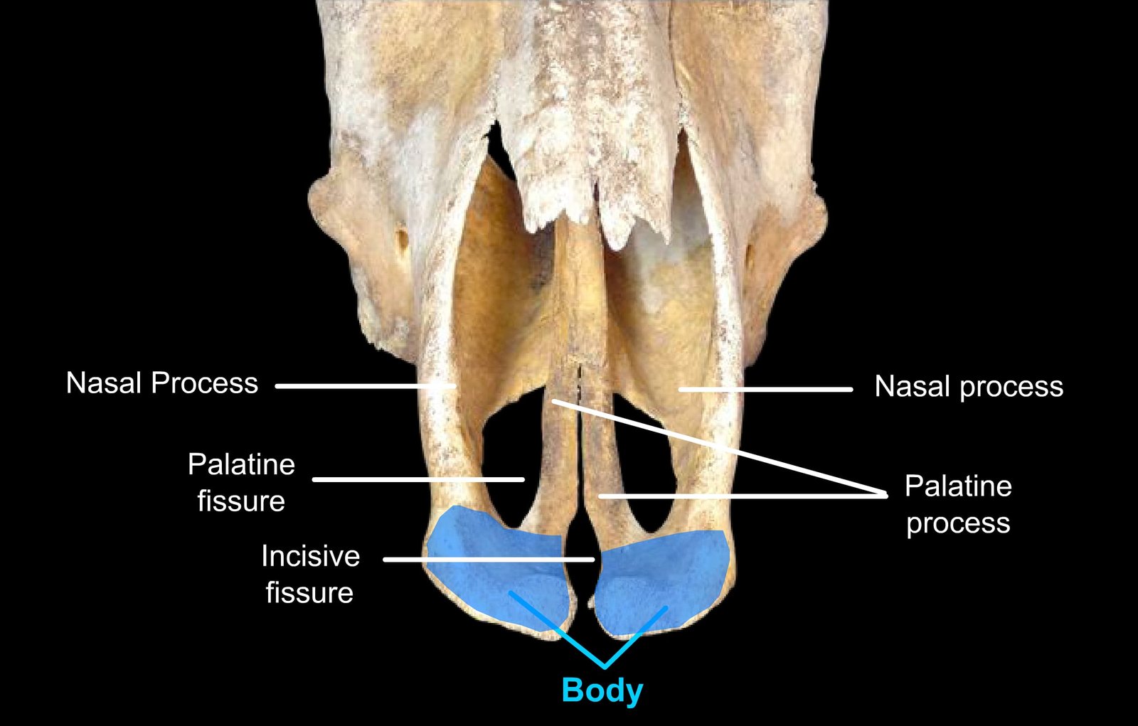
Learn the comparative anatomy of the premaxilla bone in ox, horse, dog, and fowl. Study important anatomical features, processes, foramina, and species differences in veterinary osteology.
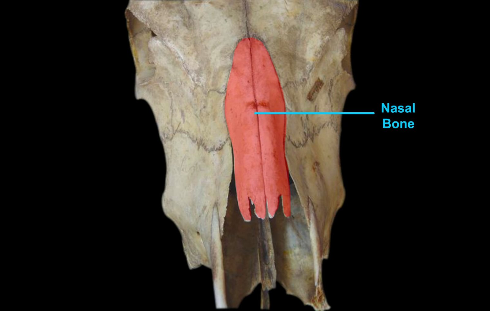
Learn the comparative anatomy of the nasal bone in ox, horse, dog, and fowl. Study important species-wise features, surfaces, processes, and veterinary anatomical differences.
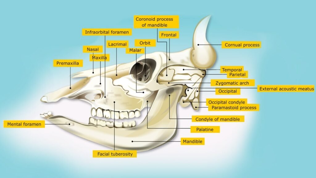
The facial bones form the framework of the face and contribute to the formation of the oral cavity, nasal cavity, orbit and jaws.