
TABLE OF CONTENTS
Sarcophaga ruficornis (Flesh Fly): Morphology, Life Cycle & Veterinary Significance
Sarcophaga ruficornis, commonly known as the flesh fly, scavenger fly, or carrion fly, is a medically and veterinary important dipteran fly associated with carrion, decaying organic matter, and animal wounds. It is a facultative ectoparasite and a producer of myiasis, with its larvae infesting wounds and feeding primarily on necrotic tissue. Due to its ability to cause traumatic myiasis and delay wound healing in livestock and companion animals, Sarcophaga ruficornis is of considerable veterinary importance.
Morphology
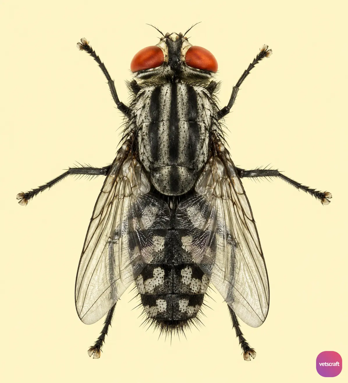
- The body is grayish-black in color, and the eyes are red.
- The arista is bilaterally plumose up to the middle, while the remaining portion is bare.
- The thorax has three longitudinal dark stripes.
- The dorsum of the abdomen has chessboard-like markings.
- Both males and females are free-living, and the larvae are flesh feeders.
Life Cycle
Sarcophaga ruficornis (flesh fly) is a larviparous fly that deposits fully developed first-stage larvae (L₁) on the flesh and wounds of animals or in decomposing meat and other decaying organic matter.
Larva
- There are three larval stages, which together last for about 2 days.
- When fully developed, the third-stage larvae leave the wound, drop to the ground, and pupate in the soil for about 6 days.
- The posterior spiracles are circular and deeply seated. The outer wall has an incomplete peritreme with a distinct button. Each spiracle has three longitudinal slits directed toward the button, along with two inward projections.
Pupa
The pupa of Sarcophaga ruficornis (flesh fly) is coarctate, brown in color, and oval-shaped. The pupal stage lasts for about 6 days. The complete life cycle takes approximately 10 days.
Veterinary Significance
Sarcophaga ruficornis is an important cause of facultative traumatic myiasis in domestic animals. The larvae infest pre-existing wounds, ulcers, necrotic tissue, and soiled body regions, where they feed on dead and decaying tissue. Infestation delays wound healing, causes tissue damage, and may predispose the affected area to secondary bacterial infections.
Cases are more common in animals with neglected wounds, poor hygiene, or conditions that result in persistent moisture or tissue damage. Sheep, goats, cattle, dogs, and other mammals may be affected. Prompt wound cleaning, larval removal, appropriate wound management, and fly control are essential to prevent myiasis and reduce economic losses associated with decreased animal health and productivity.

