
TABLE OF CONTENTS
Mouthparts of Insects: Structure, Functions, Types, and Feeding Adaptations
The mouthparts of insects are highly specialized structures adapted to different feeding habits and play a vital role in their survival. Although the basic components are similar across most insects, they undergo remarkable modifications to facilitate chewing, piercing, sucking, sponging, or lapping of food.
The mouthparts of insects consist of the labrum (upper lip), which forms the upper boundary of the mouth, and the labium (lower lip), which forms the lower boundary. Between these two structures are two pairs of biting jaws: the upper pair, the mandibles, and the lower pair, the maxillae.
On the underside of the labrum, there is a small membranous structure, the epipharynx, which bears the organ of taste. These two structures are frequently fused to form the labrum-epipharynx.
On the upper surface of the labium is another membranous structure, the hypopharynx, which bears the opening of the salivary duct. Both the maxillae and the labium possess jointed palps that are sensory in function.
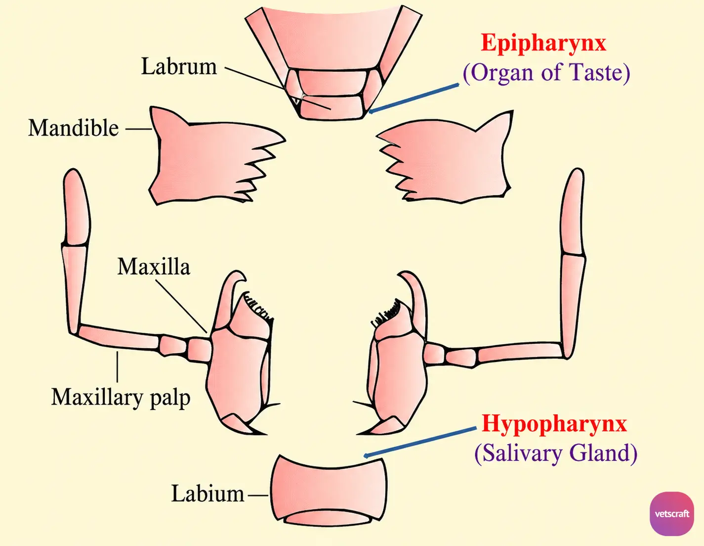
These mouthparts may be modified according to the feeding habit. The labium may be greatly expanded for imbibing liquid food (e.g. housefly), or all mouthparts may be modified into fine piercing stylets (e.g. mosquitoes).
Functions of Mouthparts
- Labrum: Draws food toward the mandibles and protects the anterior mouthparts.
- Mandibles: Cut, crush, and masticate food.
- Maxillae: Assist in mastication through forward and backward movements.
- Labium: Forms either a supporting sheath or part of the piercing mouthparts.
- Epipharynx: Organ of taste.
- Hypopharynx (Lingua/Tongue): Conducts salivary secretions.
- Maxillary Palps: Assist in sensory perception and food handling.
- Labial Palps: Function like hands to grasp food and clean the antennae, forelegs, and mouth region. They are sensory in nature and also help clean the mouthparts.
Types of Mouthparts
1. Based on Structure
- Prognathous: Projecting forward (e.g. lice)
- Hypognathous: Directed vertically to the body axis (e.g. mosquitoes, muscid flies)
- Opisthognathous: Projecting backward (e.g. bed bugs)
2. Based on Feeding Habit
- Lapping, Sponging, or Sucking Type
- Piercing and Sucking Type
- Cutting and Sponging Type
Lapping, Sponging, or Sucking Type (e.g. housefly and most cyclorrhaphan flies)
The lapping, sponging, or sucking type of mouthparts is divided into three parts: the rostrum, haustellum, and labella.
- Rostrum: It belongs to the head proper and is shaped like an inverted pyramid attached anteriorly to the clypeus above the labrum. The posterior side contains the pharyngeal tubes, and the maxillary palps are attached to its anterolateral aspect.
- Haustellum: It is the continuation of the rostrum and consists of the labrum-epipharynx, hypopharynx, and labium. The mandibles and maxillae are absent. The anterior labrum-epipharynx and posterior hypopharynx together form the food channel, which joins the outer end of the pharynx.
- Labella: The distal end of the labium terminates in a pair of oval lobes called the labella. Each labellum contains numerous pseudotracheae, which are chitinous food channels that direct food into the mouth opening of the food canal. The labellar lobes are expandable and can be molded to the feeding surface.
- In some muscid flies (e.g. Stomoxys), several rows of prestomal teeth are present between adjacent rows of pseudotracheae. These teeth help pierce the skin.
Piercing and Sucking Type (e.g. mosquitoes, Phlebotomus, Culicoides, fleas, and sucking lice)
The mouthparts consist of a lanceolate hypopharynx and a pair of mandibles and maxillae modified into stylets for piercing the skin. These structures are enclosed by the labium, which forms the proboscis sheath. The powerful suction generated by the pharynx enables blood to be drawn from the host.
Cutting and Sponging Type (e.g. Tabanus fly)
The mandibles and maxillae are elongated and pointed, functioning as stylets to pierce the skin. Once the capillary network is pierced, blood is released.
The blood is then sponged or sucked into the labium with the help of the pseudotracheal membrane and passes into the mouth through a specialized food channel located between the labrum and hypopharynx.

