
TABLE OF CONTENTS
External Anatomy of Insects: Head, Compound Eyes, Ocelli & Antennae
The insect head is a highly specialized region that houses the primary sensory organs and mouthparts. It contains important structures such as the compound eyes, simple eyes (ocelli), antennae, and associated sclerites, all of which play essential roles in vision, sensory perception, feeding, and species identification.
Head
The head is an ovoid or globular capsule composed of a number of plates or sclerites. The dorsal wall is formed by the vertex, and the lateral wall by the genae.
The anterior wall of the capsule is formed by the frons (forehead), which articulates dorsally with the vertex and ventrally with the clypeus. The incomplete posterior wall is known as the occiput, which articulates with the neck.
The head has a pair of compound eyes, which are placed laterally above the cheeks (genae). There is a central median suture that divides into two frontal sutures. Just below the frontal sutures is a hardened plate known as the clypeus, and anterior to the clypeus is the upper lip (labrum).
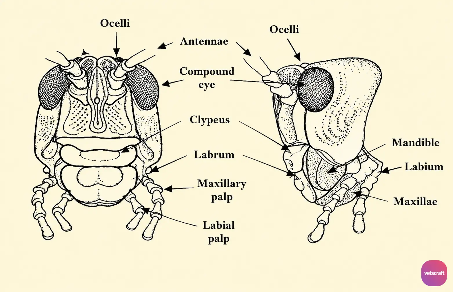
On the ventral side is the other lip, the labium. Between the labrum and labium are the mouthparts, and on either side of the labrum are the maxillary palps.
1. Compound Eye
The compound eyes are made up of individual visual units consisting of a large number of hexagonal-shaped facets termed ommatidia (singular: ommatidium), which are the basic units of the compound eye. These ommatidia contain rods and cones that aid in vision.
- Holoptic Eyes: The compound eyes are very close to each other or may meet at the midline (e.g., male insects).
- Dichoptic Eyes: The compound eyes are widely separated (e.g., female insects).
2. Simple Eyes or Ocelli (Singular: Ocellus; Maximum Number: 3)
These are photoreceptors (not used for vision) arranged in an inverted triangle on the dorsum (vertex). They are dark, hemispherical structures that project above the general level of the head.
3. Antenna
All insects have filamentous sensory organs called antennae (absent in arachnids). They are segmented structures inserted between the eyes and vary among species.
The antennae are long and slender in Nematocera (e.g., mosquitoes) and have many segments (up to 16 segments, e.g., Phlebotomus), whereas in Cyclorrhapha, the antennae have three segments (e.g., Musca and Stomoxys).
A typical antenna consists of three main segments. The first segment, attached to the head capsule, is called the scape and is generally larger than the other segments.
The second segment is the pedicel, which contains an auditory structure called the Organ of Johnston. The third segment is collectively called the flagellum, which is elongated and composed of multiple subsegments.
Types of Antennae
There are 16 types of antennae that help in the identification of insects. The most common types found in insects of veterinary importance are:
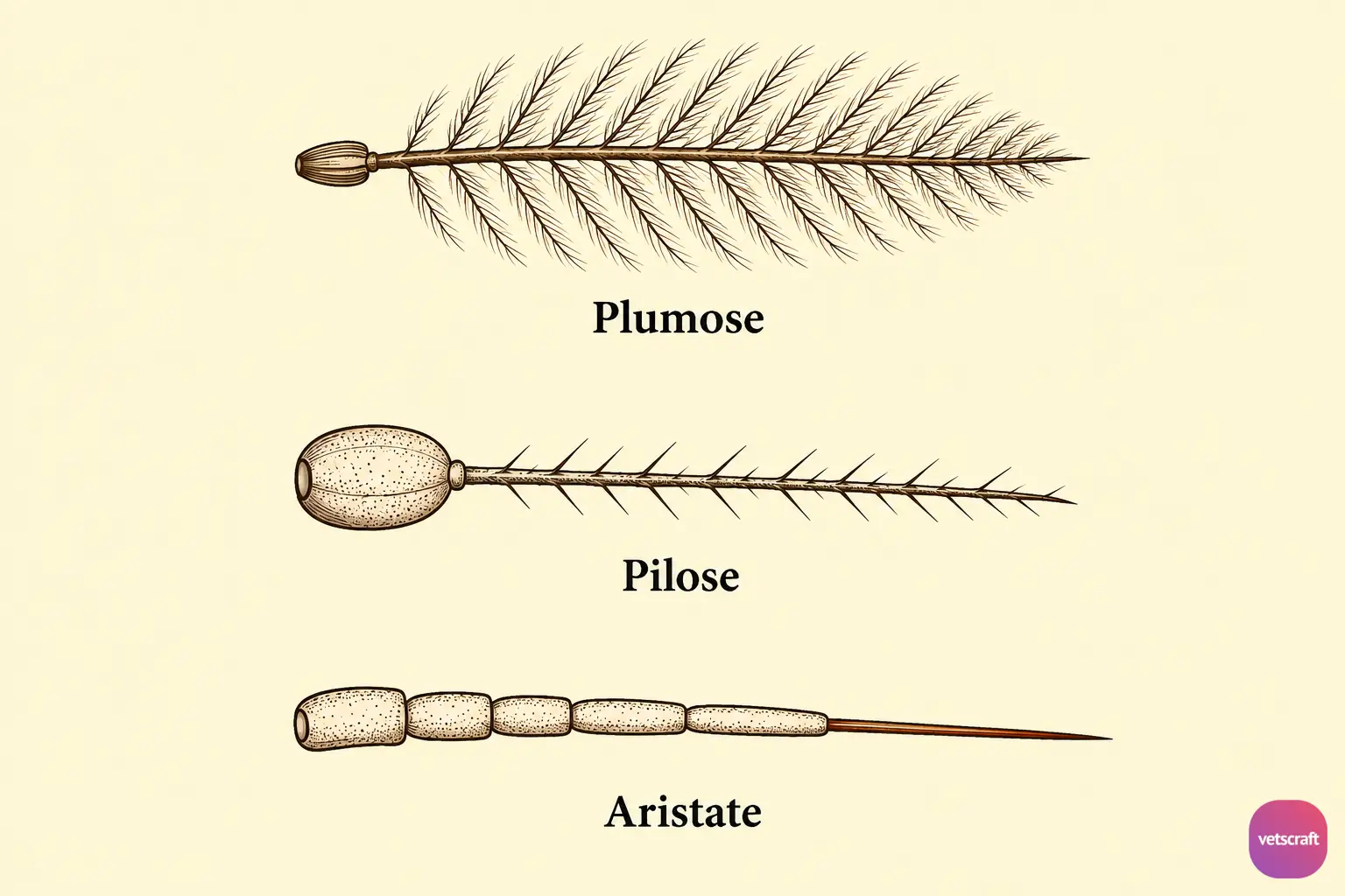
- Plumose Type: Antennae bearing a large number of bristles are called plumose antennae (e.g., male mosquitoes).
- Pilose Type: Antennae bearing a few bristles are known as pilose antennae (e.g., female mosquitoes).
- Aristate Type: A specialized bristle on the third antennal segment is called an arista, which has a sensory function (e.g., Musca spp.).
Functions of Antennae
- Antennae have sensory and tactile functions.
- Antennae serve as organs of smell and hearing.

