
Tick Morphology: Morphological Characters, Body Regions, Mouthparts, Legs, and Identification Features
Ticks are important ectoparasites of veterinary and medical significance and belong to the class Arachnida and order Ixodida. They are broadly classified into hard ticks and soft ticks based on the presence or absence of a scutum. Their morphological features, including the capitulum, mouthparts, scutum, legs, eyes, festoons, spiracles, and genital openings, are important for identification and classification.
General Morphology
Ticks are comparatively large and flattened dorsoventrally. They have a chitinous shield or plate covering the body. Those with this chitinous plate or shield are known as hard ticks, while those without it are known as soft ticks. This chitinous plate or shield is known as the scutum.
Thus, ticks with a scutum are known as scutate ticks or hard ticks, while those without a scutum are known as soft ticks or nonscutate ticks.
In male hard ticks, the scutum covers the entire body, while in females, the scutum covers approximately one-third of the body. In larvae and nymphs of hard ticks, the scutum is confined to the anterior aspect.
Body Regions
The body of the tick is oval in shape and is divided into the gnathosoma and idiosoma. A notch present at the anterior end of the idiosoma is known as the camerostoma, into which the capitulum is inserted.
Rostrum and Mouthparts
Hard ticks are broadly classified based on the length of their rostrum into two groups. Ticks with a short rostrum are known as brevirostrate, while those with a long rostrum are known as longirostrate. In hard ticks, the mouthparts project forward, while in soft ticks, the mouthparts are placed ventrally and are not visible dorsally.
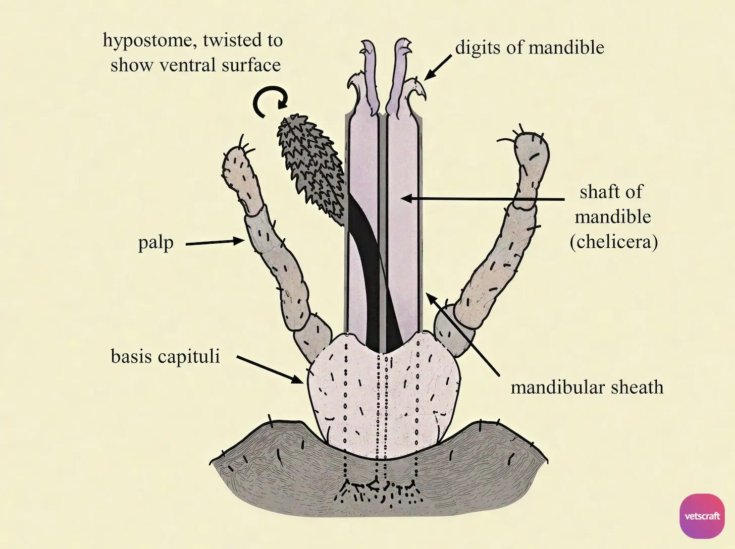
The rostrum consists of a pair of pedipalps, which are jointed structures. They are sensory in function. Internal to the pedipalps is a pair of structures known as chelicerae. Each chelicera has a base and shaft and ends in a pair of articulated digits bearing teeth.
Four teeth are seen at the tip of the chelicerae, by means of which ticks tear the skin of the host. Behind the chelicerae, there is a club-shaped structure known as the hypostome, which consists of two portions fused together and is armed with backwardly directed spines that help anchor the tick to the host’s skin.
Gené’s Glands and Porose Areas
The dorsal aspect of female hard ticks has two circular depressions called porose areas, which are the openings of Gené’s glands. Secretions from Gené’s glands provide a coating for the eggs.
Scutum and Ornamentation
When the scutum of hard ticks shows ornamentation, such ticks are known as ornate ticks. Those that lack ornamentation are known as inornate ticks.
Similarly, some soft ticks have disk-like areas on the body known as mammillations.
Eyes and Festoons
Eyes may be present or absent. In hard ticks, eyes are present on the dorsal aspect at the level of the second pair of legs. Some ticks lack eyes. In soft ticks, two pairs of eyes may be seen ventrally on the supracoxal fold, between the first and second pairs of legs and between the second and third pairs of legs.
The posterior margin of the body of ticks may have a number of folds called festoons. They are usually 11 in number. In some species of ticks, festoons are absent. There may also be a number of grooves on the dorsal aspect, such as the cervical groove, lateral groove, posterolateral groove, and posteromedial groove.
In female hard ticks, Gené’s glands, or cephalic glands, open dorsally between the camerostoma and basis capituli.
The eggs are rolled in the secretion of Gené’s glands. Hence, the eggs are seen on the dorsal aspect. These glands secrete a wax-like substance that provides a coating for the eggs.
Legs and Haller’s Organ
On the ventral aspect, four pairs of legs are seen on the idiosoma. The legs are seven-segmented, namely, coxa, trochanter, prefemur, femur, tibia, protarsus, and tarsus. The tarsus always ends in claws.
The shape of the coxa is important in the identification of ticks. The dorsal aspect of the tarsus of the first pair of legs has a sensory structure known as Haller’s organ, which helps the tick locate its host.
Spiracles and Genital Openings
In hard ticks, a pair of spiracles are seen behind the last coxa on the ventral aspect. In soft ticks, spiracles are seen between the third and fourth coxae.
In female ticks, the genital opening is seen as a transverse opening between the second coxae and is called the tocostome. In males, it is located at the same place but is smaller and is known as the epiandrium.
The anal opening is seen as a longitudinal opening posteriorly. In addition, there is an opening between the first and second coxae known as the coxal gland opening.
Ventral Plates in Male Hard Ticks
The ventral aspect of male hard ticks has a number of plates or shields called ventral plates. In female hard ticks, ventral plates are absent.
They are helpful in the identification of ticks. The plate anterior to the genital opening is the pregenital plate, and the plate immediately posterior to the genital opening is the median plate. The plates on the sides are called epimeral plates.
A groove seen in front of the anal opening is known as the anal groove. Around the anus, there is an anal plate, behind which is the adanal plate. Adjacent to the adanal plate, there is an accessory adanal plate, and below these plates, there is a subanal plate.

