
TABLE OF CONTENTS
Gasterophilus in Horses: Morphology, Life Cycle, Pathogenesis, Diagnosis & Treatment
Gasterophilus spp., commonly known as horse bot flies, are important parasitic flies that primarily affect horses and other equines worldwide. Their larvae develop in the oral cavity and stomach, where they can cause irritation, ulceration, colic, and other gastrointestinal lesions. Understanding the morphology, biology, life cycle, pathogenesis, diagnosis, treatment, and control of Gasterophilus is essential for veterinary students and practitioners involved in equine health management.
The genus Gasterophilus includes several species of veterinary importance that affect equines. The following are the major species along with their common names:
- Gasterophilus intestinalis (Gasterophilus equi): Armed horse bot fly, stomach bot fly, nit fly
- Gasterophilus nasalis: Throat bot fly
- Gasterophilus haemorrhoidalis: Nose bot fly, hip bot fly, rectal bot fly
Gasterophilus belongs to the series Schizophora, section Acalypterae, and family Gasterophilidae.
Morphology
Gasterophilus flies are large, about 18 mm long, dark brown in color, with the body covered with yellowish hairs. The mouthparts are reduced and functionless.
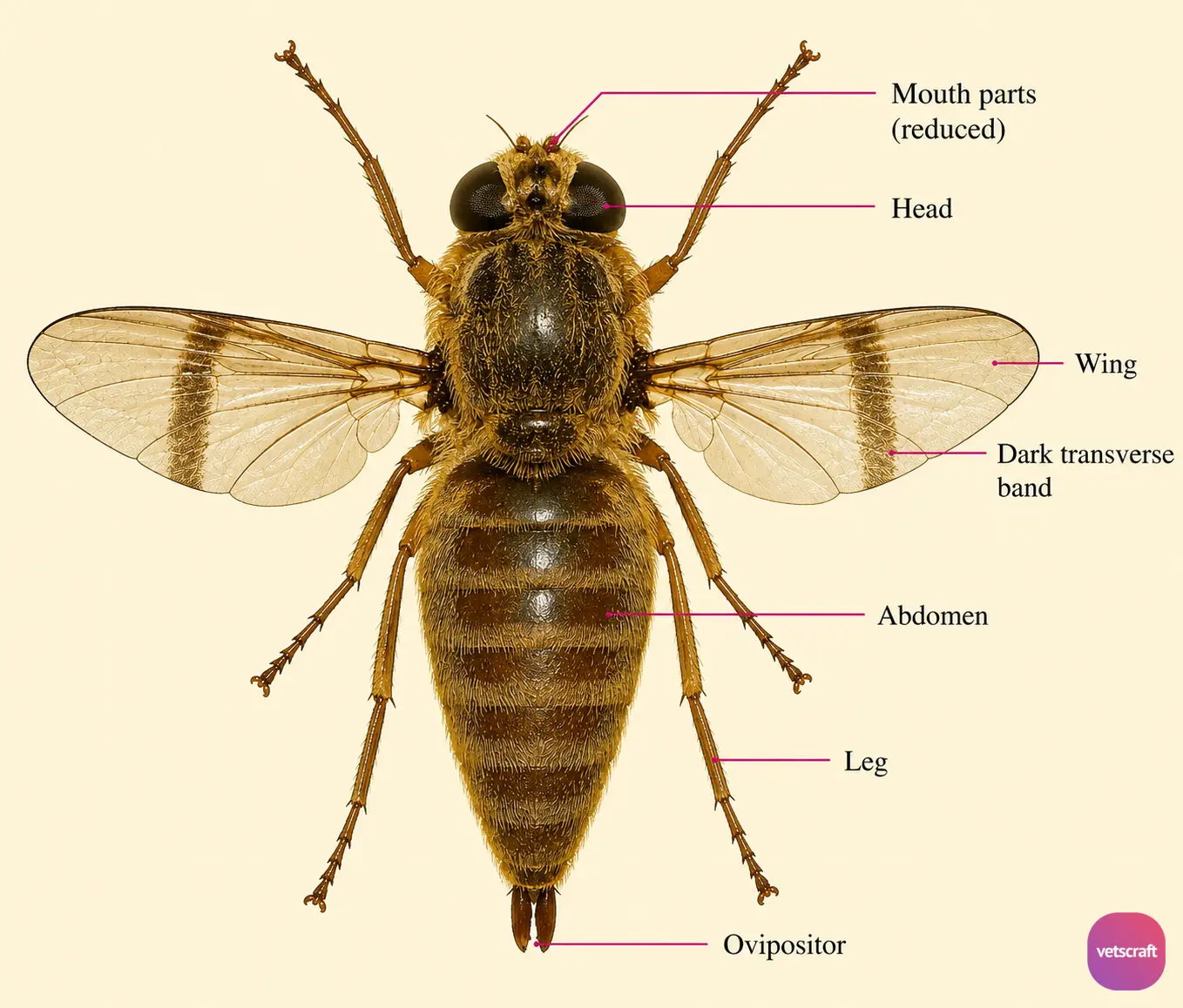
In the female, the ovipositor is long and curved. An irregular transverse band runs across each wing. Females are autogenous. The larvae feed on tissue exudates.
Biology
Larvae of this genus are found in the stomach of horses and are known as “bots.” They are host-specific. The female fly hovers above the animal with its ovipositor extended, and oviposition occurs in flight. The eggs are attached along the distal half of the hairs.
Large numbers of eggs are found mainly around the fetlock of the forelimb and also in the scapular region.
Life Cycle
The yellow-colored eggs hatch in 5–10 days, but development is greatly delayed in cool weather. Hatching occurs only with the application of moisture and friction, which are provided by the host’s rubbing and licking behavior.
The larvae are not swallowed directly into the stomach but instead penetrate the oral mucosa and gradually migrate through the mucosa as far as the pharynx before eventually reaching the stomach. Once inside the mouth, the larvae penetrate the dorsal mucosa of the anterior aspect of the tongue and burrow slowly toward the posterior end.
The larvae remain in the tongue for at least 24–28 days. The first molt occurs in the tongue, and the second-instar larvae attach to the pharynx and epiglottis for a few days before passing into the cardiac portion of the stomach. They do not undergo another molt until 5 weeks have elapsed.
Third-stage larvae are generally found in clusters in the nonglandular part of the stomach. They possess oral hooks and two rows of body spines. The larvae remain in the host for about 10 months.
Posterior spiracles are bean-shaped and have three wavy spiracular slits.
Pathogenesis
- Annoyance, colic, and perforation of the stomach and small intestinal wall.
- Ulceration of the esophageal region and stomach is the most common lesion.
- Sometimes the larvae are closely packed, producing a “palisade effect“.
- Since the majority of the larvae are found in the nonglandular region, there are usually no significant pathogenic effects. They may cause dermatitis and inflammation of the pharynx, esophagus, stomach, or rectum. Sometimes rectal prolapse may occur. Large numbers of larvae in the pyloric region may interfere with sphincter function.
- Occasionally, abscess formation, gastric rupture leading to peritonitis, or pyloric stricture due to clusters of larvae and tissue reaction in the pyloric region may occur.
- Occasionally, larvae may penetrate human skin and cause “creeping myiasis.”
Diagnosis
- Eggs can be found by examining the knees, pasterns, and fetlocks.
- Larvae in the pharynx can be seen by direct inspection.
Treatment
- Carbon disulfide is administered by stomach tube or in a capsule at a dose of 2–5 mL/kg body weight after an 18-hour fast.
- A piperazine/carbon disulfide complex (Parvex) or this mixture plus phenothiazine (Parvex Plus) at a dose of 110 mg/kg is effective.
- Trichlorfon is administered in feed or water at a dose of 39.6 mg/kg body weight.
- Thiabendazole/trichlorfon is administered in feed or as a drench at a dose of 2 g thiabendazole and 18 g trichlorfon per 50 kg body weight.
- Dichlorvos in feed at a dose of 26–52 mg/kg or as a paste at 20 mg/kg is effective.
- Ivermectin can be administered at a dose of 200 µg/kg.
- Treatment against Gasterophilus should be carried out once or twice per year.
- Purgatives such as oil of chenopodium, turpentine, and linseed oil have also been used to remove larvae from the stomach.
Control
- Periodic grooming of horses removes the eggs before they become infective to the host.
- A warm water insecticide wash (40–43°C) can be sprayed on areas containing eggs.
- Warm water stimulates egg hatching, and the insecticide kills the larvae.

