
TABLE OF CONTENTS
Chrysomya (Blow Fly) in Veterinary Parasitology: Morphology, Life Cycle & Pathogenesis
Chrysomya genus flies, also called beauty-bearing flies, belong to the family Calliphoridae. These flies are of major importance in veterinary parasitology because certain species are obligate ectoparasites and cause myiasis in animals. Among them, Chrysomya bezziana, commonly known as the Old World screwworm fly, is the most important veterinary species due to its ability to invade living tissues.
In veterinary parasitology, understanding the morphology, life cycle, and pathogenesis of Chrysomya species is essential for the diagnosis, treatment, prevention, and control of myiasis. This article covers the important species of Chrysomya, their identifying morphological features, life cycle, larval characteristics, and the pathological changes associated with infestation.
The Chrysomya genus has two important species:
- Chrysomya bezziana (Blue-green bottle fly, Old World screwworm fly)
- Chrysomya megacephala (Oriental latrine fly)
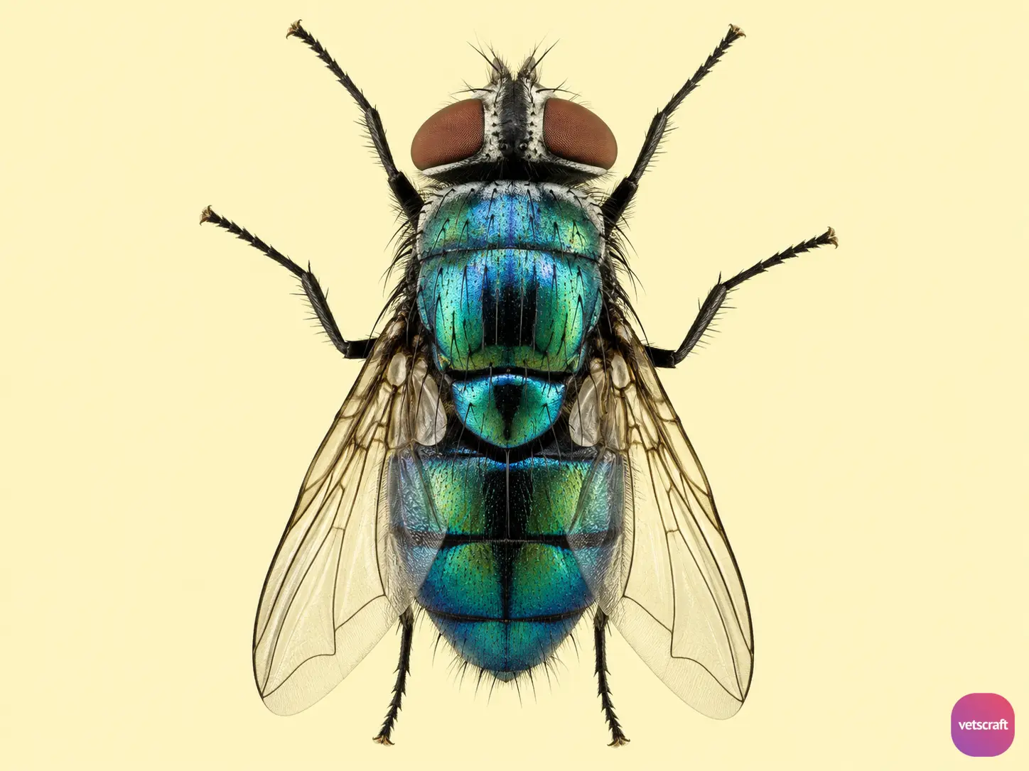
Calliphorid flies are commonly known as blow flies, screwworm flies, or maggot flies.
Morphology
- The face is pale-colored, and the body is green or blue with narrow dark bands along the posterior margin of the abdominal segments.
- The thorax has two faint dark stripes, and the legs are dark.
- Both males and females are free-living, and the larvae are flesh feeders.
Life Cycle
Female flies are autogenous. Eggs are laid by the female over pre-existing wounds or in body orifices such as the eyes, ears, or nostrils.
About 175–200 eggs are laid at a time. The eggs hatch within 12–18 hours, and the larvae emerge.
Larva
- There are three larval stages. The L₁ larvae migrate to the wound or moist tissue, where they aggregate and feed on wound fluids.
- During the 6–7 days of feeding, the larvae burrow deeply into the host tissues so that only the posterior segment and spiracles remain exposed.
- When mature, the third-stage larvae leave the wound, drop to the ground, and pupate in the soil for about 12 days.
- The larvae are long, maggot-like, white in color, 12-segmented, and have transverse rows of backward-directed spines, giving them the name “screwworm” larvae. The larvae are carnivorous and feed on the larvae of primary myiasis flies.
- The posterior spiracle is located at the posterior end of the larva and is oval or circular in shape. The outer wall has an incomplete peritreme. Each spiracle has three longitudinal spiracular slits without a button.
Pupa
The pupa of Chrysomya is coarctate, brown in color, and oval-shaped. The pupal stage lasts for about 8 days. The entire life cycle takes approximately 15–20 days.
Adult
Chrysomya adults are diurnal, and both males and females are free-living.
Pathogenesis
Chrysomya causes intermittent irritation and pyrexia, followed by the formation of cavernous lesions. The affected tissue undergoes progressive liquefaction, necrosis, and hemorrhage.
Histologically, two phases are observed. The first phase is characterized by intense neutrophil infiltration and hemorrhage associated with larval growth. The second is a fibroplastic healing phase in which mast cells and eosinophils are prominent.

