Histology of Penis

Veterinary Anatomy

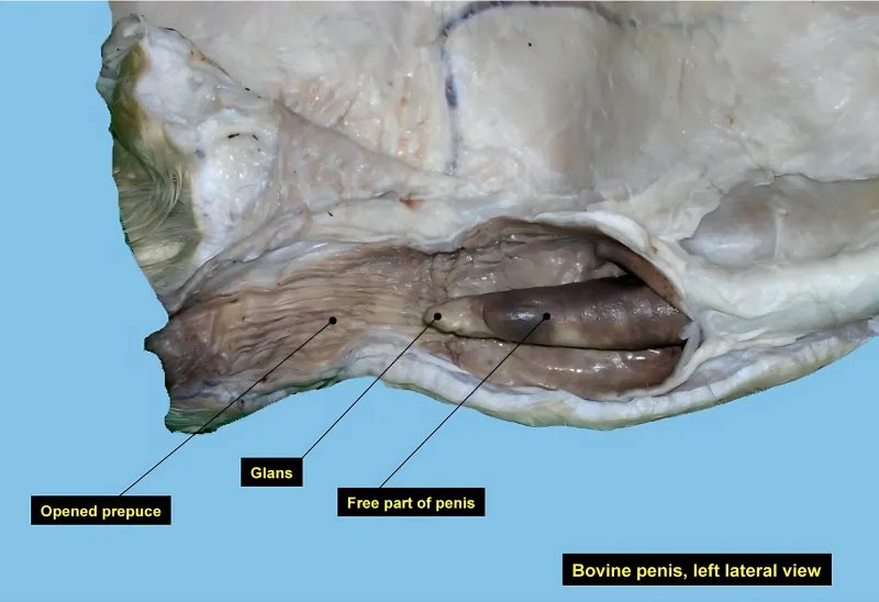
Penis consists of a body and glands. Body consists of The erectile corpus cavernosum penis, whose two crura are attached to the ischial arch. Corpus cavermosum urethrae or corpus spongiosum penis surrounding urethra muscles. Glans penis forms the tip of penis. Body of penis Consists of the corpora cavernosa, which have a capsule, a system of trabeculae and […]

Histology of Penis Read More »

Histology of Vas deferens

Veterinary Anatomy

Ductus deferens or Vas deferens has a mucosa, muscularis and fibrosa. The mucosa form longitudinal folds and is lined by pseudostratified columnar cells similar to that of ductus epididymis. But the cells are low and stereocilia shows variable distribution, absent form some cells and present in others. Lamina propria is rich in elastic tissue. Muscularis

Histology of Vas deferens Read More »

Histology of Epididymis

Veterinary Anatomy

Histology of Epididymis Epididymis is a highly convoluted tube to form a solid body so that in section the tube is cut several times. The duct is enveloped by collagenous tunic, which contains circular smooth muscle. The duct has a wide lumen and the mucosa has no folds. The epithelium is pseudostratified columnar ciliated and

Histology of Epididymis Read More »

Histology of Respiratory division of Lungs

Veterinary Anatomy

From the respiratory bronchioles onwards, the structures contain alveoli in their walls and serve both as conducting and respiratory passages. The epithelium is initially simple cuboidal but become flattened or squamous later. No Goblet cells or glands occur in any part. The respiratory structures consist;  Respiratory bronchiole,  Alveolar duct,  Alveolar Sac,  Alveoli. Respiratory bronchiole These

Histology of Respiratory division of Lungs Read More »

Histology of Lungs

Veterinary Anatomy

The lungs are paired organs located in the thoracic cavity. The surface of each lung is covered by a serous membrane (pleura). Each lung is divided into a number of lobes and pleura, dips into interlobar fissures tissue and covers interlobar surfaces. Each lobe is sub-divided by interlobular tissue into lobules. Each lobule is pyramidal

Histology of Lungs Read More »

Scroll to Top