TABLE OF CONTENTS
Reproductive Abnormalities in Cows and Heifers
To identify reproductive tract abnormalities in cows and heifers, we have to go through history of animal, physical examination, vaginoscopy, rectal examination, etc.
Reproductive abnormalities in cows and heifers is examined by:
- History
- Vaginal Examination
- Rectal Examination
- Laparoscopy
- Ultrasonographic Examination
History
History taking can be done simultaneously while the animal is being examined. The important issues that need to be addressed include the following:
- Parity (virgin heifer, pregnant heifer, uniparous or multiparous cow).
- Age (including age at first calving).
- Cyclic history (normal or abnormal cycle lengths, anestrus, nymphomania).
- Calving dates and comments (dystocia, twins, retained placenta, surgical or mechanical intervention, viability of calf).
- Breeding dates and methods (artificial insemination or natural service, estrus detection methods and personnel, semen supplier and quality, previous record of bull fertility, including examination for venereal disease).
- Previous treatments (drugs, dosages and routes, treatment intervals, clinical outcome, drug withdrawal disease).
- Nutritional program (periparturient supplementation of beef cows, dry-period feeding of dairy cows, body conditions of cows at calving, milk production levels).
Physical Examination
Visual inspection gives valuable information about the individual animal and will be an aid to genital examination.
Visual inspection involves the following:
- General conformation
- Conformation of the external genitalia
- Vulvar discharges
- Condition of the mammary gland & Udder
- General behaviour of the animal
1. General Conformation
Cows with a masculine appearance of the head and shoulder region may be suffering from Cystic ovarian degeneration Freemartin heifers may exhibit steer-like appearance.
2. Conformation of the External Genitalia
Physiological Alterations
- The vulval labia are normally covered with soft, thin skin and are symmetrical and closely opposed to ensure closure of the vestibule and vagina.
- There is high tonicity of the pelvic diaphragm as well as the vulva in a non-pregnant or early pregnant cow manifested by firmness of the diaphragm and relatively small and wrinkled appearance of the vulval lips.
- During the last trimester of pregnancy these structures undergo gradual but continuous relaxation.
- The relaxation becomes marked and the vulva nearly doubles in size during the last 10 to 14 days prior to parturition and this extreme relaxation of the pelvic ligaments causes the tail head to become elevated.
- Within 2 weeks following parturition the vulva returns to normal size.
Pathological Alterations
Pathological alterations involve the following:
- Extreme relaxation of the pelvic diaphragm, enlargement of the vulva and elevation of the tail head in Cystic ovarian degeneration
- Relaxation of the vulval lips alone in cows with long standing cases of metritis.
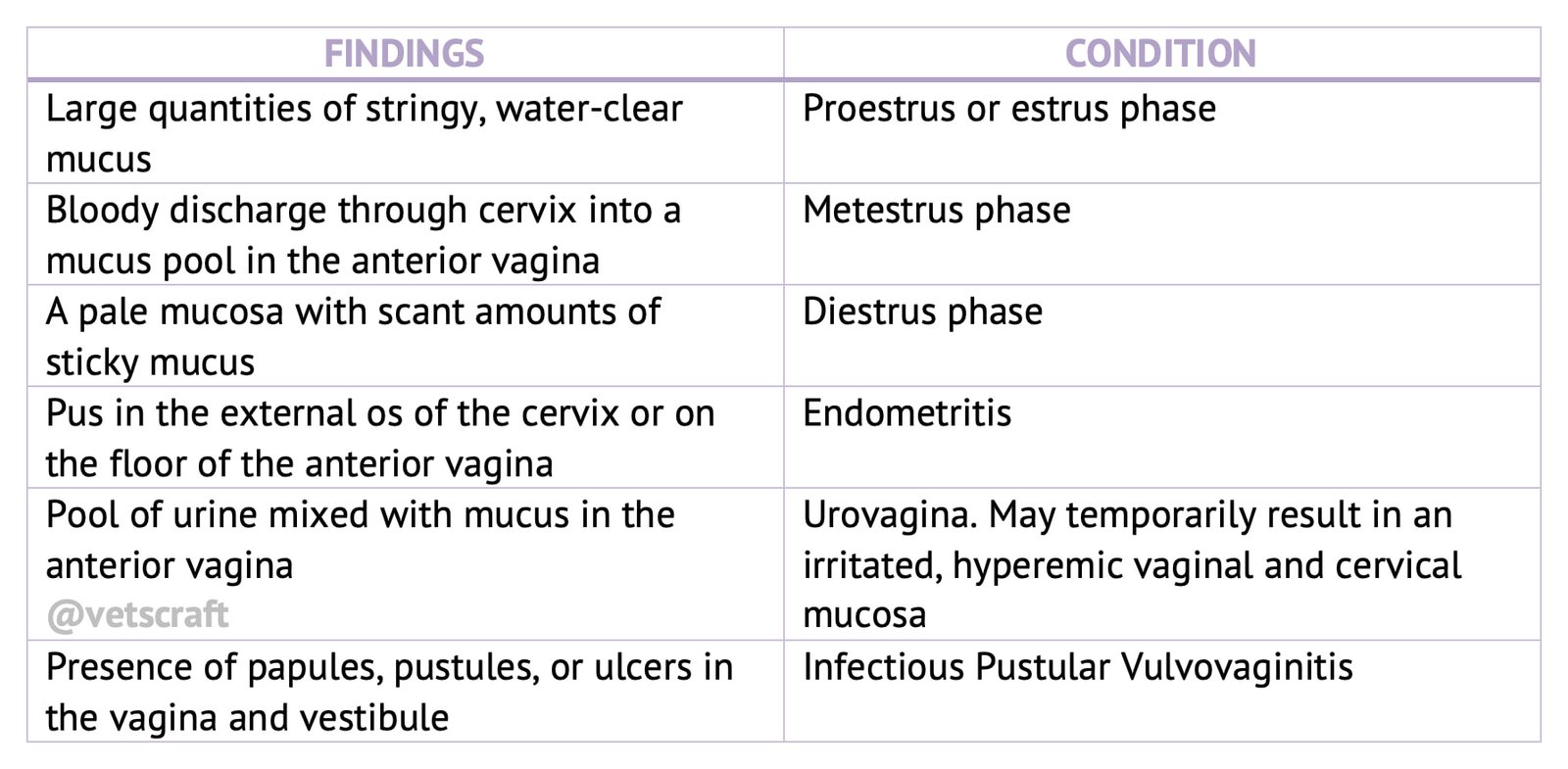
- Extreme swelling of the vulva associated with edema, but with increased tension, is found as the first sign of Infectious Pustular Vulvovaginitis (IPV).
3. Discharges from the Vulva
Discharges Observed in Normal Animals
During estrus, the vulva may appear edematous with presence of characteristic clear, elastic mucus that hangs from the ventral commissure. In many cases the mucus may be present adhering to the tail.
Blood stained mucus may be present in some heifers and cows during the first few days after the end of estrus referred to as metestrual bleeding.
Reddish grey discharge, consisting of blood elements and debris of endometrium referred to as lochia, is observed in post parturient cows and increases in amount reaching peak quantities around the third week of the post partum period.
Discharges Associated with Pathological Conditions
The presence of a mucopurulent (clear mucus discharge with pus flakes) to purulent discharge indicates:
- inflammation of any segment of the reproductive tract.
- infection of the urinary system.
It should be noted that apparent purulent discharge may be absent in cases of pyometra while a heavy purulent discharge may be present in animals with normally progressing pregnancy.
Greyish discharge not containing apparent pus has been observed in certain cases of cystic ovaries.
4. Condition of the Mammary gland & Udder
- Edema and enlargement of the mammary gland are normally found in the pre parturient and post parturient period.
- Cows that have failed to conceive over long periods may have a small shrunken vulva.
5. General behaviour of the Animal
The general behaviour of the animal can be observed only when the animal is not confined. Signs of estrus, hyper-estrus, bellowing and pawing can be observed.
Vaginal Examination
Supplemental information obtained by vaginal examination helps to refine the tentative diagnosis made after rectal examination of postpartum cows. However, it is seldom employed in the cow.
Manual Examination
Manual examination of the vagina and cervix of the early postpartum cow will aid in the diagnosis of the following conditions:
- Vaginal/cervical trauma.
- Retention of fetal membranes.
- Patency of the cervical canal.
The cow‘s vulva and perineum should be carefully washed with a mild disinfectant soap, and a lubricated disposable plastic sleeve should be worn by the examiner to perform vaginal examinations.
Vaginoscopic Examination
After washing the vulva and perineum, the speculum is inserted first in a dorsalcranial direction until the ischial symphysis has been passed, then in a cranial direction.
Slight resistance will be noticed at the vestibulovaginal junction which can be easily overcome by gentle pressure.
With a torch light, the vaginal vault should be examined for:
- the location of the cervix,
- cervical or vaginal color and secretions,
- cervical anomalies,
- trauma and discharges.
Rectal Examination
Rectal examination involves:
- Examination for pregnancy
- Examination for non pregnancy
Examination for Pregnancy
Pregnancy diagnosis is based on detection of the physiological changes of the genital organs associated with pregnancy.
The uterus is the organ mainly involved and the positive signs of pregnancy include:
- Palpation of amniotic vesicle
- Palpation of fetal membrane slip
- Palpation of placentomes
- Palpation of fetus
Examination of Normal Non-pregnant Reproductive Tract
The size, muscular tone and contents of the uterus should be assessed. This can be done simultaneously with the “membrane slip” for pregnancy determination. Commonly used terms for characterizing uterine tone include the following:
- Estrus tone: a turgid, contracted uterus that is often curled into a rather tight configuration.
- Diestrus (“normal”): a relaxed muscular uterus.
- Edematous: a somewhat turgid uterus but without muscular contraction; may be palpable for a few days after estrus.
- Flaccid: a limp, soft, usually thin-walled uterus that does not contract in response to palpation.
- Thickened (“doughy”): a pathologic description, indicating thickening of the endometrium and possibly the myometrium as well.
- Fluctuant: uterus in which there is intra luminal fluid.
Post Parturient Involution
Pregnancy and involution represent the only two clinically appreciable physiological alterations of size. In pregnancy, the size progressively increases while during the involution period, the size of the uterus regresses and returns to the non pregnant state.
Palpation for Uterine Disorders
During routine post partum examinations in cases in which pregnancy diagnosis is negative or in examination of “problem cows,” the reproductive tract should be examined for palpable abnormalities.
The essential questions for the examiner to answer are the following:
- Is the uterus symmetrical and approximately the size and tone of the non gravid tract?
- Is there a corpus luteum or an ovarian follicle associated with corpus luteum or an ovarian follicle associated with increased uterine tone that is indicative of cyclicity?
- Are there any palpable lesions of the reproductive tract?
Uterine Inflammation
It is generally possible to diagnose moderate to severe endometritis, acute metritis or pyometra by rectal examination.
Adhesions, Abscesses and Tumors
Adhesions, abscesses, and tumors of the uterus are significant pathological conditions that can impair reproductive performance in female animals.
Fetal Remnants
- Occasionally, a fragment of an autolyzed term fetus may remain in the uterine lumen following parturition
- Can be detected as a moveable firm mass in the lumen of an involuting uterus. A foul vaginal discharge will often be noted.
- Cows or heifers that do not calve at the expected time following a positive pregnancy diagnosis may have either a mummified or macerated fetus.
- In cases of fetal maceration, a distended uterus with palpably crepitant fetal bones can be felt. An ipsilateral CL may be present, as well as a fetid vaginal discharge.
- The prognosis for future fertility of such cows is grave due to severe damage to the endometrium .
Freemartinism
In Freemartinism the cervix is rudimentary while the uterus is underdeveloped and characterized by the presence of two thin walled, very narrow tubes occupying the sites of the normal horns, suspended in ligamentous sheets resembling the broad ligaments.
Lateral exploration along the edge of the broad ligaments leads to location of barely perceptible thickening indicating the rudimentary ovary.
Failure to locate the normal cervix during the course of rectal examination should always be followed by a thorough exploration for signs of freemartinism.
White Heifer Disease
White heifer disease also known as segmental aplasia of the Mullerian duct. The extent of aplasia and the number of the missing segments is variable
Secretion of the normal segments becomes entrapped between the missing segments or anterior to the missing part, resulting in marked distension of the normal segment associated with thinning of the wall. Persistence of the “hymen,” one of the forms of white heifer disease, results in accumulation of secretion in the anterior part of the vagina, with consequent dilation which elicits tenesmus.
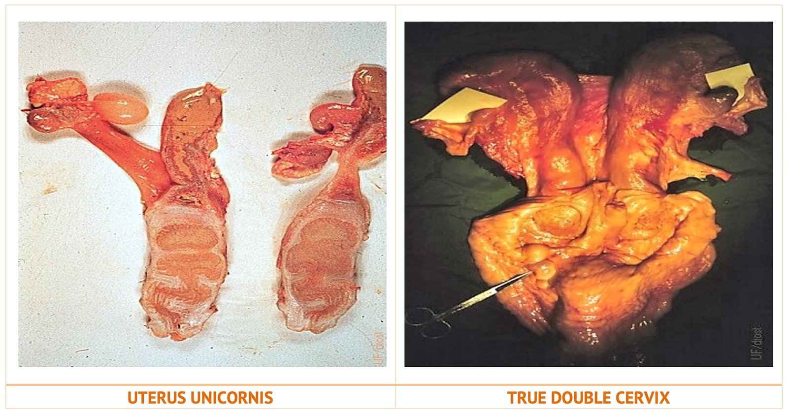
Uterus Unicornis
Uterus unicornis relatively rare abnormality has been found in practically all breeds. The horn that is present is functionally normal and conception is possible only during an estrus when the follicle develops and ovulates occurs in the ovary on the side of the normal horn
Reduced fertility can be anticipated.
Cervix Duplex or Double Cervix
Cervix duplex or double cervix also is a rare abnormality. The presence of two cervices, resulting in two single tube genital tracts anterior to the vagina, might cause temporary confusion in the examiner‘s mind. Diagnosis, however, is easy.
Fertility of the affected animal does not have to be impaired if natural breeding is employed. Artificial insemination might result in failure to conceive unless the ovary carrying the follicle ready to ovulate is detected and the semen is deposited in the cervix of the same side.
Bilateral insemination in the double cervices might also be recommended.
Pyometra
Pyometra is characterised by accumulation of pus in the uterus and may occur due to lack of sufficient relaxation of the cervix or to the presence of cervicitis combined with atony of the uterus and consequent lack of expulsive force.
The amount of exudates varies from 25cc., which is barely felt upon examination, to several liters.
The uterine walls are thinner than in the non-pregnant uterus, but thicker than the pregnant uterus The condition has to be differentially diagnosed from pregnancy.
Mucometra or Hydrometra
Both mucometra and hydrometra are similar except for the degree of hydration of mucin present in the uterus which may vary from a watery fluid to a semisolid mass.
Mucometra or Hydrometra condition is observed in heifers or cows following:
- Arrest in the development of mullerian duct system
- Persistence of hymen
- Prolonged hormonal stimulation with estrogens or progestogens
Cows with mucometra or hydrometra due to defects of genital tract are sterile. Cows with pyometra do not cycle, while cows with a hydrometra do.
Chronic Nonproductive Metritis
Chronic nonproductive metritis condition is often referred to as chronic endometritis
On rectal palpation:
- The uterus lacks tone,
- Has a thin wall, especially in the intercaruncular spaces,
- Caruncles, therefore, appear more prominent, and the endometrial surface feels wavy and uneven.
Diagnosis is by vaginal examination and histological examination of biopsies.
Abnormalities of the Ovaries
Common abnormal conditions of ovaries include smooth ovaries, ovarian cysts, ovarian hypoplasia, underdevelopment of ovaries in heifers, ovaritis or oophoritis, and miscellaneous conditions such as abscesses and tumors.
Abnormalities of Oviduct
Hydrosalpinx and pyosalpinx are reproductive pathological conditions affecting the oviduct (fallopian tube) in female animals.
Abnormalities Involving Mesosalphinx and Ovarion Bursa
Clinical differentiation between parasalpingitis, perisalpingitis and ovarian bursitis is practically impossible.

For clinical purposes, perisalpingitis appears to be the most correct term to describe the inflammation and the consequent thickening and adhesion formation involving mesosalpinx, mesovarium and salpinx. Other structures in the area, such as the ovaries, the horns of the uterus and others, might also be embedded in the adhesions.
Very fine adhesions between the ovary and fimbria-the fringes of the edge of the infundibulum-are present in numerous animals, especially immediately after ovulation. These do not appear to interfere with the normal function of the oviduct.
Laparoscopy
The reproductive tract can be directly visualized by laparoscopy or endoscopy.
Ultrasonographic Examination
The uterus and ovaries can be indirectly examined by ultrasonographic techniques
Real-time ultrasound, in which a two-dimensional “sonic picture” is generated from echoes
Ultrasonography used to diagnose pregnancy, normal ovarian structures, uterine and ovarian pathology, etc.

