TABLE OF CONTENTS
Echinococcosis
Echinococcosis is a parasitic disease of animals, caused by echinococcus species.
Etiology
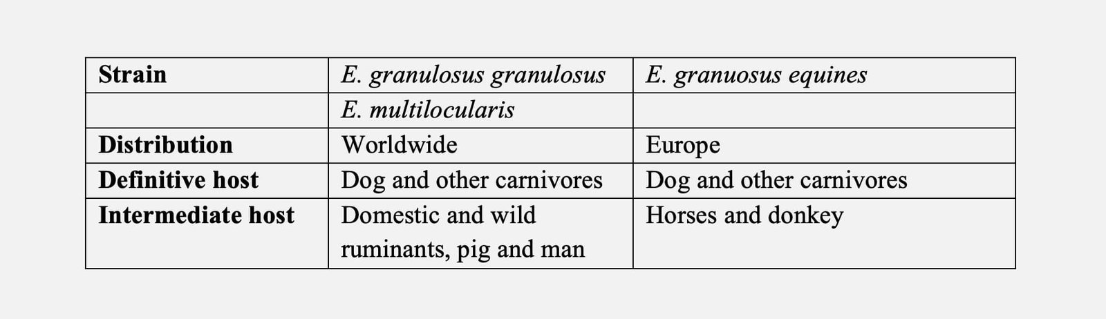
- Echinococcus granulosus (E. granulosus granulosus and E. granulosus equinus) and Echinococcus multilocularis
- Echinococcus is a smallest cestode of domestic animals (6mm), consist of 3 or 4 segments. The gravid segment is half the length of rest of segment.
Location
- Adult worm-small intestine.
- Immature/Metacestode/ hydatid stage: Lung and liver.
- Sheep-70% of cyst found in lung and 25%in liver.
- Horse and cattle: 90% of cyst found in liver.
Epidemiology
- Epidemiology of Echinococcosis is based on pastoral and sylvatic cycle.
- Pastoral cycle: The dogs are infected by feeding of ruminant offal containing hydatid cyst, which mature into adult and excrete the gravid segment through faeces. The man get infection by accidental ingestion of vegetable and other foodstuff contaminated with dog faeces.
- Sylvatic cycle: Wild canids and ruminants are involved in transmission.
Life cycle
Proglottids/egg > Intermediate host (6-12 months) > Definitive host (40-50 days).
- The prepatent period in the definite host is around 40-50 days after which only one gravid segment is shed by the tapeworm per week.
- The oncospheres are capable of prolonged survival outside the host, being viable on the ground for about two years.
- After ingestion by the intermediate host, the onchosphere penetrates the gut wall and travels in the blood to the liver or in the lymph to the lungs.
- These are the two commonest sites for larval development, but occasionally onchospheres escape into the general systemic circulalion and develop in other organs and tissues, where they develop into large, thick-walled, unilocular hydatid cysts that bud protoscolices endogenously about 6-12 months.
- Hydatid cysts have been rarely reported in the CNS of domestic animals and are rare in people, in which they produce symptoms similar to those of brain tumor.
Hydatid cyst
Types of cyst
- Sterile cyst: not infective for dog. In horse – 27% of cysts are sterile, in sheep – 51% of cyst are sterile and in cattle & pig most of cysts are sterile.
- Fertile cyst: infective for dog.
- Size: 5 – 10 cm in size (maximum of 50cm).
- Cyst wall: Outer layer is thick and made up of concentrically arranged laminated membrane. The inner layer: germinal layer and granular in nature.
- Cyst fluid: pale yellow in colour and contains 17 to 200 mg of protein per ml
- Brood capsule: protoscolices attached on the inner granular layer or detached and float free in the hydatid fluid is called as “Hydatid sand”.
- Daughter cyst: develops within the mother cyst, If mother cyst rupture release protoscolices & brood capsules and develops as external daughter cyst.
Clinical manifestation
- Dog –clinical signs are not evident.
- In domestic animals hydatid cyst in the liver or lungs is usually tolerated without any clinical signs, and the majority of infections are only revealed at the abattoir.
- Human: respiratory signs are common. Presence of several hydatid cysts in liver causes abdominal distension. If cyst rupture causes death by anaphylaxis.
Necropsy Finding
- Presence of fluid filled cyst in liver, lung and other organs.
Diagnosis
- Presence of hvdatid cyst is clinical entity rarelv suspected in domestic animals.
- Diagnosis of infection in dogs with adult tape worm is difficult, because the segments are small and are only shed sparsely.
- In some countries control regimes have involved the administration of purgative anthelmintics such as arecoline hydrochloride, so that the whole tapeworm is expelled in mucus and can be searched for in the faeces.
- Man: Intradermal casoni’s test.
Treatment
- Echinococcus tapeworrns are more difficult to remove compared to Taenia sp.
- Praziquantel is highly effective @ 5.0-12.5 mg/kg b.wt. After treatment it is advisable to confine dogs for 48 hours to facilitate the collection and disposal of infected faeces.
- Man: surgical excision of cyst.
- Aspiration of cyst fluid and irrigation of cyst cavity with 2.5 % – 10% formalin (not commonly used) or own serum. Care should be taken to avoid spillage of cyst fluid, while aspiration.
- High dose of mebendazole, albendazole and praziquantel may be effective.
Control
- Regular anthelmintic treatment for adult tape worm.
- Avoid feeding of cyst containing offals.
- Proper disposal of sheep carcass in the farm.
- Denying dog access to abattoir.
E. multilocuaris
- DH: Wild canids, domestic dog and cat
- Location: Small intestine
- IH: Rodents, Large mammals and man
- Metacestode stage: Hydatid cyst in liver is mostly multilocule
Pathogenesis
Egg > IMH > small intestine > Release of oncosphere > Development of adult metacestode stage > Liver and lung > systemic circulation.
Clinical signs
- Clinical signs based on severity of infection and location of cyst.
- Heavy infection causes impairment of affected organs functions.
- Rupture of cyst is leads to anaphylatic shock.
- Alveolar cyst, a diffuse growth with many compartment containing gelatinous matrix into which scolices budding off.
Diagnosis
- Animals: rare only by post mortem examination.
- Man: Serology : CFT and CIE.
Treatment
- Aspiration of cyst fluid and irrigation of cyst cavity with 2.5 % – 10% formalin (not commonly used) or own serum. Care should be taken to avoid spillage of cyst fluid while aspiration.
- Surgical removal of cyst.
- Higher doses of mebandazloe, albendazole and praziquantel.
Control
- Similar to T. ovis.

