TABLE OF CONTENTS
Toxocariasis
Toxocariasis is one of the largest nematodes disease in domestic animals. Presence of adult worm in the intestine may cause unthriftiness and occasionally intestinal obstruction.
Etiology

Morphology
- Large white worm.
- The egg is dark brown and subglobular, with a thick pitted shell.
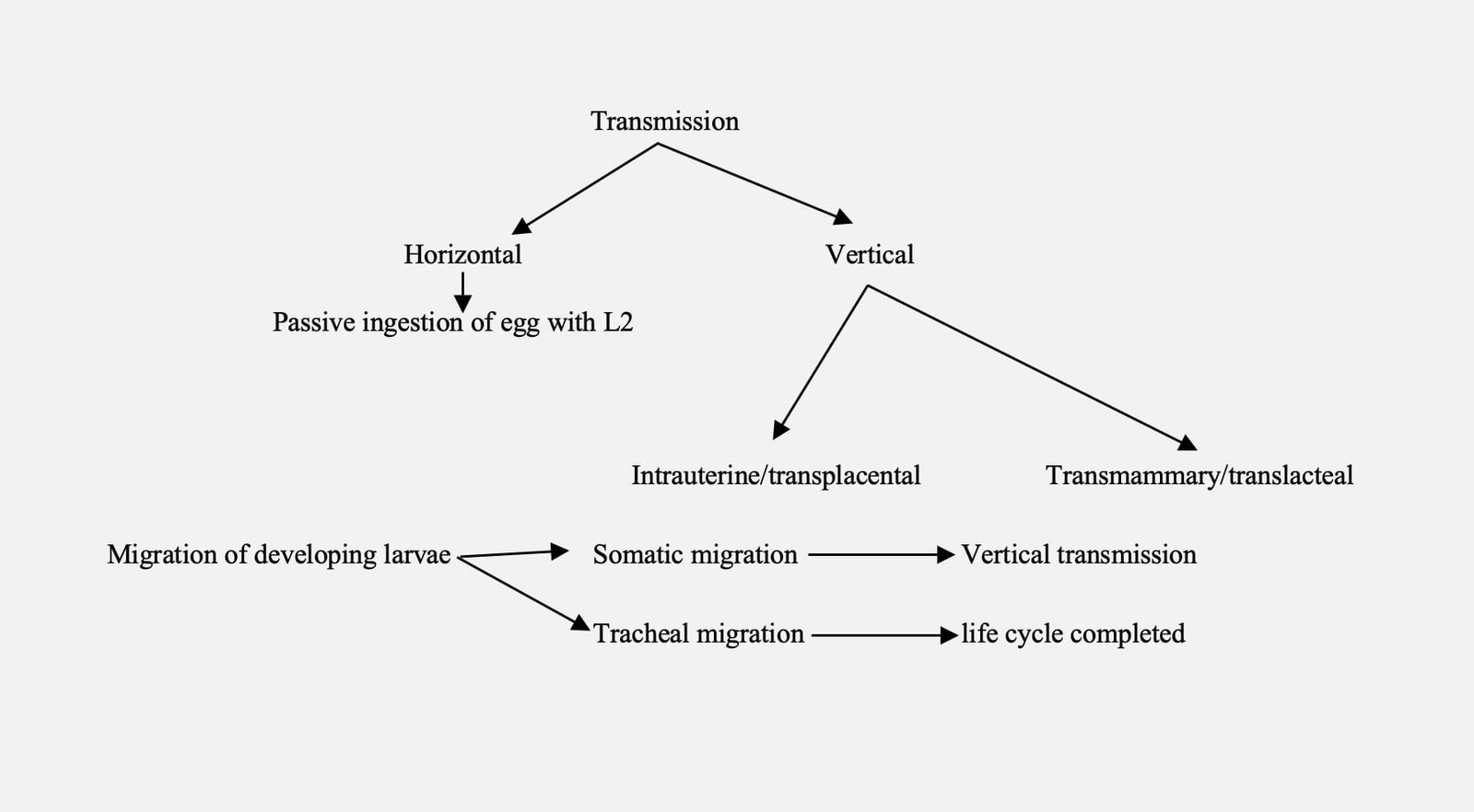
Transmission and life cycle
Transmission and life cycle of toxocara is direct and complex in nature.
Toxocara vitulorum in Bovines
Toxocara vitulorum is the largest intestinal parasite of cattle.
Host affected
- Cattle, buffalo, sheep and goat
- Buffalo calves are highly susceptible due to prenatal and neonatal infection
- Larvae present in dams milk up to 30 days after parturition
- If no tissue migration – prepatent period: 3 – 4 weeks
- Pregnant cows body tissues acts as a reservoir of larvae for its offspring (features is the reservoir of larvae in the tissues of cows with subsequent milk borne transmission, ensuring that calves are exposed to infection from the first day of life).

Life Cycle

Pathogenesis
- Light infection – less than 70 larvae / calf – infection unnoticed.
- Heavy infection – 70 -500 larvae / calf – clinical disease.
- Lung: Verminous pneumonia – haemorrhagic & necrotic foci.
Clinical signs
- Intermittent diarrhoea, steatorrhoea and mud coloured evil smelling faeces, pot-belly appearance and poor thriving.
- Colic signs resembling intestinal obstruction (may be piling of adult worm in intestine).
- Calf hood mortality in buffalo calves.
Diagnosis
- Based on history and clinical signs.
- Identification of characteristic eggs.
Treatment
- Benzimidazole compounds-fenbendazole and oxfendazole can be used.
- Morantel citrate – 10 mg / kg -mature and immature worm.
- Levamisole – 7.5 mg / kg – mature and immature worm.
- Piperazine – 250 mg / kg b.wt.
Control
- The prevalence of infection can be drastically reduced by treating calves at 3rd and 6th weeks of age to prevent the developing worms reaching patency.
Prophylactic Schedule (Buffalo calf)
- First dose – 15th days of age.
- Second dose – 30th days of age.
- Third dose – 3rd month of age.
- Fourth dose – 6th month of age.
- Fifth dose – one year of age.
Toxocara canis
- Host: Dog and fox
- Location: Small intestine
- Morphology: similar to Toxocara vitulorum.
Epidemiology
- Puppies less than six months are highly susceptible
- Paratenic host: Rodents and birds ingest egg with L2, the L2 larvae settled in tissues of paratenic host.
- Definitive host pickup infection while ingestion of paratenic host infected with L2 larvae
- Recent evident that bitches may be reinfected during late pregnancy or lactation leading directly to transmammary infection of the suckling pups
- Somatic tissues of bitch constant reservoir of infection for puppies as well as adults and unsusceptible to most anthelmintics.
Prepatent period
- Ingestion of eggs with L2 without somatic migration: 3- 4 weeks.
- Ingestion of paratenic host with L2: 4 –5 weeks.
- In puppies due to prenatal infection: 3 weeks.
Transmission

Life cycle
Life cycle of toxocara canis is direct and complex in nature.

Weakened immunity
- Occurrence of eggs in faeces of bitches after whelping as a result of weakening of immunity and contaminated environment.
- Lactogenic hormone (prolactin), which permits mobilization of larvae from somatic tissues to small intestine through lung, larvae undergo maturation and become adult.
- Habit of whelped bitches to eat faeces of puppies, which may contains immature worms. The immature worms enter small intestine of bitches and become adult.
- Post parturient infection are eliminated within a few weeks of the termination of lactation.
- Not all larvae are mobilized during pregnancy and some may remain dormant in tissues and eliminated through subsequent pregnancies.
- The duration of larvae in bitches may be long and animals infected for upto 385 days and is being capable of transmitting infection to puppies.
- Factors which induce mobilization and migration are unclear but probably there is a hormonal basis involved.
Pathogenesis
- In moderate infections, the larval migratory phase is accomplished without any apparent damage to the tissues.
- In heavy infection the pulmonary phase of larval migration is associated with pneumonia. This is sometimes accompanied by pulmonary oedema. The adult worms cause mucoid enteritis; there may be partial or complete occlusion of the gut and in rare cases, perforation with peritonitis or in some instances blockage of the bile duct.
- Aberrant site
- Bile duct-inflammatory changes and occlusion.
- CNS-Nervous disorder.
Clinical manifestation
- In mild to moderate infections, there are no clinical signs during the pulmonary phase of larval migration.
- In heavy infection migratory larvae causes pneumonic signs such as coughing, increased respiratory rate and frothy nasal discharge.
- Heavy infection in puppies leads to death of whole litters.
- Most fatalities from T. canis infection occur during pulmonary phase and pups which have been heavily infected transplacentally and may die within a few days of birth.
- The adults in the intestine may cause pot-belly, with failure to thrive, and occasional diarrhoea. Entire worms are sometimes vomited or passed in the faeces.
- Aberrant site in CNS – convulsion (yet to be proved).
- Death due to intestinal obstruction.
Diagnosis
- Based on history and clinical signs.
- Demonstration of eggs on direct smear method/Centrifugal sedimentation/Flotation method.
- Excretion of adult worm in the faeces.
Treatment
- In dogs, compounds approved for treatment of roundworm infections include fenbendazole, milbemycin, moxidectin, piperazine, and pyrantel.
- Fenbendazole 100 mg/kg b.wt single dose or 50mg/ Kg b.wt daily for three days.
- Piperazine – mature worm – 100 mg/ kg and immature worm – 200 mg /kg.
- Pyrental pamoate – 5-10 mg/kg b.wt.
- Milbemycin oxime 0.5 mg/Kg b.wt.
- Diethyl carbamazine (DEC) – 50 mg/ kg (contraindicated for dogs with patent infection of D. immitis).
- Dichlorvos – 12 – 15 mg/ kg body weight.
- Trichlorophan – 75 mg/kg.
- Nitroscanate – 50 mg/kg.
- Mebendazole – 10 mg/ kg (twice a day for 2 days).
Control
- Good hygienic maintenance in kennels.
- The main control measures relies on reducing environmental contamination.
- Environmentally resistant larvated eggs on the ground and somatic larvae in the bitch are the main reservoirs of infection.
- Perinatal transmission of infection can be greatly reduced by treating bitches with daily doses of fenbendazole (25 mg/kg, PO) from day 40 of gestation to day 2 after whelping.
- Ivermectin (0.3 mg/kg, S/C) on days 0, 30, and 60 of gestation, and 10 days after whelping.
- Ivermectin (0.5 mg/kg) on days 38, 41, 44, and 47 of gestation, or ivermectin (1 mg/kg) on days 20 and 42 of gestation; these uses of ivermectin are extra-label.
- To minimize egg output, pups should be treated as early as possible; ideally, treatment should be given 2 wk after birth and repeated at 2-wk intervals upto 2 month of age, and then monthly upto 6 month of age.
- Nursing bitches should be treated at the same times as puppies. In cats, perinatal transmission can be greatly reduced by treating queens with a single dose of emodepside/praziquantel spot-on in the last week of pregnancy. Kittens should be treated with an appropriate anthelmintic at 3, 5, 7, and 9 wk of age, and then monthly to 6 month of age.
- Nursing queens should be treated at the same time as kittens. In other animals, the appropriate frequency of preventive treatment for roundworms should be based on a risk assessment of the animal’s environment.
Prenatal and neonatal infections of young puppies are controlled by treating of pregnant bitches and her litter with suitable prophylactic schedule as follows:

- Kennels should be made up of impervious surfaces and floor should be thoroughly cleaned
- Control the entry of rodents in the kennels
Toxocara cati
- Host: cat and wild felids.
- Location: small intestine.
- Morphology: Male – 6 cm and female – 10 cm long, Egg: Similar to T. canis ( Colourless thick pitted albuminous layer).
Life cycle
Life cycle of toxocara cati is direct.
- Prenatal infection does not occur.
- Paratenic host involves in the life cycle are earth worm, rodents, cockroaches, chicken, sheep and other animals fed with infective eggs.
- Transmammary infection only occurs.
- Larvae occur in the milk throughout lactation when queens infected with eggs and transported to the mammary gland after an extended period in other tissues before lactation commences.
Pathogenesis and clinical signs
- Pot belly appearance, diarrhoea and poor hair coat.
Diagnosis and treatment
- Similar to T. canis.
Control
- Removal of kitten from infected dams.
- Similar prophylactic schedule as that of T. Canis.
Toxocara leonina
- Host: Dog and cat
- Location: Small Intestine
- Paratenic host: Mice
- Morphology: Male – 7 cm and female – 10 cm, Egg: Slightly oval with smooth thick shell.
Life cycle
Life cycle of toxocara leonina is direct but not complex.
Egg (L2) > Passive ingestion > Small intestine > L2 > Adult > eggs.
Pathogenesis and clinical signs
- Pot bellied, intermittent diarrhoea and possibly anemia.
Diagnosis and treatment
- Similar to T. canis
Control
- Hygienic maintenance.
- Prevent the exposure.
- Prevent capturing of paratenic host.

