TABLE OF CONTENTS
Testicular Degeneration in Animals: Causes, Pathogenesis, Clinical Signs and Treatment
Testicular degeneration is a pathological condition characterized by the progressive shrinkage of the testicular tissue, leading to impaired sperm production and reduced fertility in male animals.
75 to 80 percent of testicular pathology is related to testicular degeneration including fibrosis and orchitis.
Testicular degeneration may be mild or severe and is usually bilateral as it is most commonly due to generalized disease processes.
Unilateral degeneration can occur secondary to local testicular lesions such as tumors.
Testicular degeneration may develop very rapidly within a few days or hours; while testicular regeneration proceeds slowly over weeks and months.
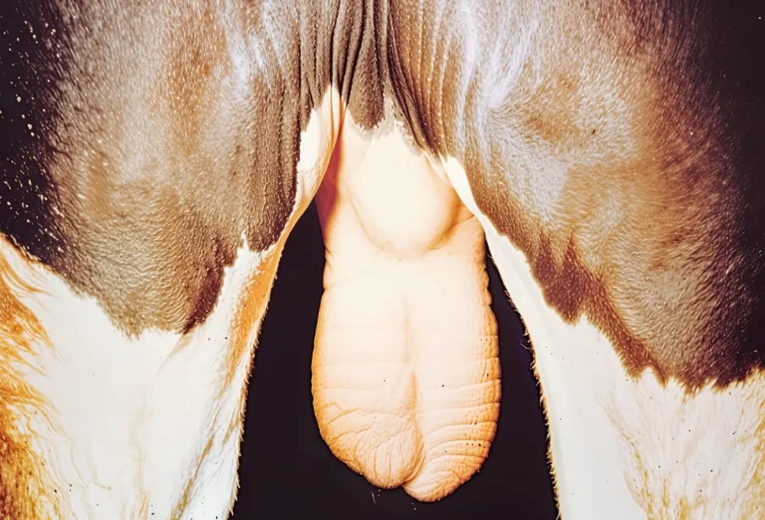
Testes with degeneration of the seminiferous tubules are usually atrophic and softer and smaller than normal testes.
In chronic cases the testicle may be firm due to fibrosis even calcium may be deposited especially in the areas just peripheral to the rete testis.
Histologically, it may be difficult to distinguish between slight degrees of testicular degeneration and hypoplasia.
Etiology
Thermal Influences
Elevation of the testicular temperature in conditions such as cryptorchid and ectopic testes; inguinal hernias; scrotal dermatitis due to, irritants, choriopic mange, myiasis in sheep, and localized skin infections or wounds; contusions and haematomas of the scrotum and testes; prolonged elevated body temperature as in certain infectious diseases and in prolonged high environmental temperatures, particularly associated with high humidity.
Testicular degeneration is most common in tropical climates and involving breeds originating in the temperate zones.
Bulls exposed to heat stress for 8 hours a day for 7 days resulted in a deleterious effect on semen quality reaching its peak at 2 to 3 weeks after the stress with recovery by 9 weeks.
Rams maintained at ambient temperatures of 900F or above develop a marked drop in semen quality with about 10 percent motility and 70 percent abnormal sperm cells within a few weeks. Recovery was not complete until 2 to 3 months after normal temperatures were restored.
High ambient temperatures will also cause lowered fertility due to testicular degeneration in boars.
Heat primarily affects the spermatids, the spermatozoa and the spermatocytes but usually does not affect the spermatogonia.
Heat has no effect on the Leydig cells.
Males that lie down for long periods of time such as bulls with bovine spastic syndrome, or males that are unable to rise, often develop testicular degeneration and atrophy due to the prolonged elevation of testicular temperature.
When the scrotal temperature of bulls was raised to 38.40C or 0.30C below body temperature, the motility and percent of live spermatozoa in the semen decreased to zero by the second week.
Damage to spermatogenic function occurred in beef bulls by low temperatures down to -250F associated with winds of 60 miles per hour causing frostbite, necrosis of skin, scrotal dermatitis, heat, swelling, testicular degeneration and adhesions.
Vascular Lesions of the Testes
Interference with the circulation and infarction of the testis can be produced by manual torsion of the testis or by the emasculatome used for castration of lambs or calves.
Congestion and pain of the scrotal testis due to naturally-occurring torsion or contusion has been reported in racing stallions. The testes would be swollen, congested and painful for the next 3 to 4 days.
Dogs with inguinal or abdominal cryptorchid testes may occasionally develop torsion of the testis with a sudden onset of pain and associated symptoms.
Hemorrhagic infarction of the testis may follow torsion.
Tumors of the abdominal testes may also predispose to torsion.
Testicular biopsies, particularly in bulls, often result in focal areas of testicular necrosis because of vessel damage when a desirable size of testicular material is obtained.
Inflammation of the testicular artery in the horse may be caused by Strongyle larvae, the equine arteritis virus and other unknown agents. This may produce areas of testicular degeneration.
Strongyle larvae can produce adhesions between the testis and its tunics.
Age associated vascular lesions such as hyaline degeneration in older bulls, rams and dogs causing degenerative changes in the seminiferous tubules.
Varicocele may affect both the circulation of blood and the heat regulatory mechanism of the pampiniform plexus. Varicoceles may be palpated in rams.
Irradiation
Irradiation produces interference with spermatogenesis by injuring spermatogonia, spermatocytes and spermatids.
The spermatocytes are most sensitive to irradiation while Leydig and Sertoli cells are quite resistant.
The amount of irradiation and length of treatment are highly important in the degree of effects produced and the rate of recovery.
Hormones
Testicular degeneration and atrophy of the testes occurs in the dog and rarely in other animals due to tumors of the anterior pituitary gland or hypothalamus interfering with the production of gonadotropic hormones. This is called Dystrophia Adiposagenitalis Syndrome in dogs.
Excessive estrogenic hormone produced by sertoli cell tumors and testosterone, and rarely estrogens, produced by Leydig cell tumors may suppress FSH production and cause testicular degeneration.
Age Effects
Permanent and progressive testicular degeneration occurs fairly frequently in all species without any indication of its pathogenesis.
Senile atrophy of the testes is common in dogs over 10 years of age and in cats over 12 years of age.
Many disease, genetic, and management factors influence this age effect.
Subacute or Acute Trauma, Stress or Disease
Subacute or acute trauma, stress or disease may cause rapid or progressive testicular degeneration in males.
Those that have been associated with reduced fertility and a decline in semen quality are: shipping under adverse conditions of heat and cold, severe fatigue, excessive physical work, traumatic gastritis in bulls, liver or abdominal abscesses, multiple severe contusions with fractured ribs, severe screw-worm infestations, moderate quittors in bulls, acute laminitis in rams, horses and other species, kickt wounds and resulting haematomas of the testes and scrotum in stallions and extensive infected wounds.
Acute or Chronic, Localized or Systemic Infectious Diseases
Acute or chronic, localized or systemic infectious diseases are common causes of testicular degeneration with moderately to severely reduced fertility in males.
Infections producing orchitis or epididymitis have a direct effect on the testes due to the inflammatory reaction causing heat, edema, congestion, circulatory interference, ischemia and even infarction due to the thick firm tunica albuginea that restricts normal swelling of the testicular parenchyma.
In bacterial diseases localizing in the testes, abscessation may occur.
Infectious agents resulting in orchitis are Brucella abortus, both field strains and strain 19 in bulls, Brucella suis in boars miliary or chronic tubercular infections with Mycobacterium tuberculosis of the testes in bulls and boars, Corynebacterium pyogenes, in bulls and rams, Actinomyces bovis in bulls, Malleomyces mallei in horses; Salmonella abortus equi and “epizootic cellulitis” due to the arteritis and the influenza viruses in horses; Corynebacterium ovis and Pasteurella pseudotuberculosis in rams; lumpy skin disease in cattle; sheep pox virus in rams, IBR-IPV virus, that markedly affected the spermatocytes and caused arrested spermatogenesis in bulls.
Brucella canis in dogs caused scrotal swelling, epididymitis and unilateral or bilateral testicular degeneration, fibrosis and sterility.
“EPIVAG” also causes an orchitis with testicular degeneration and atrophy.
Orchitis in rams from which a P.L.T. agent (chlamydia) was recovered.
Sporadic infections of the testis with staphylococci, streptococci, E.coli, proteus and Pseudomonas organisms have been reported as a cause of orchitis in dogs and other male domestic animals.
Systemic Infectious Diseases
Pneumonia and shipping fever, I.B.R.-I.P.V., equine infectious anemia, actinobacillosis, strangles, blue-tongue virus vaccination, tick-borne fever infection in rams.
Those infectious diseases causing gradual debilitation, loss of weight and testicular degeneration with atrophy include: foot and mouth disease, actinomycosis, Johne’s disease, pyelonephritis, chronic peritonitis, and lymphomatosis.
Nutritional Factors
Diets for males sufficient for growth and maintenance are adequate for fertility. Testicular degeneration is caused by under-feeding or starvation.
Chronic diseases associated with inanition and debility and testicular atrophy may include, chronic severe parasitisms, either external or internal such as: lice, mange, ticks, roundworms, flukes, and etc., senile changes due to worn or diseased teeth, chronic arthritis, tumors, fat necrosis around the large intestine in bulls, severe spastic syndrome and other possible diseases interfering with the intake and passage of food through the gut.
Malnutrition may complicate these other chronic diseases.
Improper care and management of bulls accustomed to special care and feeding may cause a marked loss in weight resulting in impotency and testicular degeneration and atrophy.
Severe vitamin A deficiency characterized by night blindness, lacrimation and opthalmia usually precedes testicular degeneration and poor semen quality.
Malnutrition, debility and cachexia produce degeneration and atrophy of the seminiferous epithelium by causing a suppression of the release of gonadotropic hormones from the pituitary.
A high level of feeding and the accompanying obesity has no effect on semen quality in a normal male, but does effect their libido and willingness to breed.
Poisons or Toxins
Poisons or toxins may adversely affect the germinal epithelium. Testicular degeneration in bulls and rams with hyperkeratosis caused by the chlorinated naphthalenes.
Antimony compounds injected for the treatment of heartworms in dogs have been reported to cause temporary sterility.
Chemicals, metals and earth salts produce degeneration of the seminiferous epithelium in a variety of species. eg. alkylating agents, cadmium chloride and amphotericin B.
Auto-immunization
Auto-immunization may play a role in a few cases of testicular degeneration. Degeneration has been produced experimentally in rams, bulls and laboratory animals by the subcutaneous injection of autologous testicular material, together with Freund’s adjuvant. Degeneration may occur with or without inflammation.
No natural cases of testicular degeneration due to auto-immunization have been reported.
Testicular Neoplasms
Testicular tumors are unusual in most domestic animals except the dog and possibly old bulls. Primary testicular tumors are usually of three main types and originate from the interstitial cells, the sertoli cells and the germinal epithelium.
This may be due to a genetic predisposition in dogs and to the maintenances of intact male dogs until they become senile.
Large tumors result in testicular degeneration probably due to the compressing effect of the tumor on the adjacent seminiferous tubules.
The degeneration may also be due to the excess steroids produced by the interstitial, or sertoli cell tumors.
Testicular neoplasms in various animal species:
- Bulls: Testicular tumors are seldom observed except in old animals. Interstitial cell tumors of a benign type are occasionally observed in bulls over 7 to 10 years of age. These may be multiple and in some cases they are large enough to be palpable as small, round, masses, liver-like in consistency and causing the testes to feel lumpy on palpation.
- Stallions: Testicular tumors have been described only occasionally. Rare dermoid cysts or teratomas of the testicle of the horse are often found in cryptorchid testes.
- Rams: Testicular tumors are rare.
- Boars: Testicular tumors are very unusual.
- Cats: Testicular tumors apparently are rare.
Genetic or Hereditary Factors
Acquired testicular degeneration have been reported in identical twin bulls. Genetic constitution appeared to influence the onset of generalized arthritis, fat necrosis, traumatic gastritis and associated testicular degeneration in identical twin bulls.
Some bulls can more resistant to testicular generation than other bulls when affected by similar etiologic agents.
Symptoms and Signs
They are similar in all species of animals and range in degree from mild to severe depending upon the cause and duration of the degeneration.
Fertility
In the male with testicular degeneration may vary from slightly reduced conception rates to moderate to severe infertility to complete sterility.
Size of the Testes
Size of the testes is usually reduced from normal to about one-half to two-thirds of its normal size depending on the duration and degree of atrophy of the seminiferous tubular epithelium.
In acute orchitis, inflammatory conditions, obstruction of the efferent ducts, or testicular tumors, an increase in testicular size is usually noted.
Consistency of the Testes
Consistency of the testes is soft and flabby in acute moderate to severe testicular degeneration.
In mild testicular degeneration consistency differs only slightly from an elastic, tonic, slightly tense, fluctuating normal consistency.
The tonometry is useful for the objective measurement of testicular consistency. The tonometer test correlated closely with other tests of semen quality.
Bulls with soft testes tended to produce fewer sperm cells.
Acute orchitis is characterized by a tense swelling and enlargement of the testis accompanied by pain and heat. This should be differentiated from hydrocele or hemaotcele.
Chronic degeneration and atrophy of the testes especially in older males is characterized by fibrosis of the testes often with calcification. Fibrotic testes are hard, firm and often lack tonicity or elasticity.
Ultrasonics can be of value in the detection of connective tissue in fibrosis of the testes.
Testes containing tumors may be enlarged and have an irregular shape and consistency on palpation.
Libido or Sex Drive
Libido or sex drive is usually not related or associated with testicular degeneration except in acute orchitis or other painful testicular diseases or in testicular atrophy associated with severe debility or inanition due to a variety of causes.
Leydig cells are much more resistant to stress factors than the cells of the germinal epithelium.
Semen Picture
Semen Picture is very helpful in diagnosing testicular degeneration. Semen may be collected by the artificial vagina, by electroejaculation or other methods.
Since it requires about 60 days or more for sperm cells to develop from spermatogonia until they are ejaculated, it may be desirable in diagnosing testicular degeneration to take several samples at weekly or greater intervals.
The semen production and quality in young immature bulls continues to improve in motility, concentration and morphology for months after puberty is reached so if an initial semen sample is poor, subsequent samples are indicated.
The first semen samples taken after a long period of sexual rest may also be misleading as these are often of poor quality with many dead spermatozoa.
In males where testicular degeneration is severe, semen quality is usually poor.
Sperm Cell Concentration
Sperm cell concentration may be reduced to one-third to one-half of normal values or show severe oligospermia or azoospermia. Watery translucent semen is common in affected bulls and rams.
Sperm Cell Motility
Sperm cell motility is reduced one-third to one-half or more due to an increase in abnormal cells, dead cells, necrospermia, or poorly viable cells. The duration of motility after ejaculation is shortened.
Sperm Cell Morphology
After staining it reveals an increase in abnormal heads, tails and middle pieces. The degree of change in the morphology of cells may vary from normal semen which usually contains from 5 to 15 percent abnormal cells, to a 15 to 20 percent increase in abnormal cells, to a 15 to 20 percent increases in abnormal cells in mild cases of testicular degeneration, to a 20 to 30 percent increase in moderate testicular degeneration, and to a 35 to 60 percent or greater increase in severe testicular degeneration.
The occurrence of large numbers of primary abnormalities that arise during spermatocytogenesis or spermatogenesis and early spermiogenesis are more indicative of a severe testicular degeneration than the presence of secondary abnormalities that occur late in spermiogenesis or during storage of spermatozoa in the epididymis.
The appearance of primitive cells such as giant cells and spermatocytes with restitution or pyknotic nuclei together with a marked reduction in sperm cell concentration and motility are signs of severe degeneration of the testes.
Testicular biopsies may be of value in certain species such as the dog and horse but are impractical for technical reasons in the bull.
Furthermore the above signs and additional tests of sequential semen samples will accurately reveal the relative degree of testicular degeneration.
Prognosis
The prognosis in testicular degeneration is variable depending upon the causative factors, the duration and degree of the degeneration and the age and value of the male.
Although testicular degeneration can occur rapidly within a few hours, days or a week or more, recovery is very slow usually requiring 3 to 6 months or more in moderate to severe cases.
The prognosis in slight or mild case of testicular degeneration in young or middle aged bulls due to transient and correctable causes is fair to good.
Controlled breeding during the recovery period may result in fair to good fertility in some males.
The prognosis is guarded to fair in young males with only moderate evidence of testicular degeneration, in older males suddenly developing infertility due to a transient disease where advanced testicular changes do not occur; in mild cases of trauma to the testes.
The prognosis is poor in progressive senile testicular degeneration, in acute severe orchitis especially if it is bilateral and associated with abscessation, in advanced fibrosis and atrophy of the testes, in degeneration associated with hypoplasia in young to middle-aged males, and in older males with bilateral tumors of the testes.
Treatment
The treatment of testicular degeneration requires the correction or the alleviation of the causative factors.
Sexual rest is usually advised, since in most case, fertility is reduced to a point where the use of the male is questionable.
Males with mild degrees of degeneration not severely influencing fertility should be used sparingly so as to maintain as large number of normal, motile spermatozoa in each ejaculate.
A balanced ration high in vitamin A and possibly a good quality and variety of protein are indicated.
Good quality roughage or pasture is highly desirable. Some exercise is usually recommended for the larger farm animals.
If excessive heat and humidity is the cause of the temporary infertility in bulls, air conditioning or cooling is indicated.
Hormones are of no proven value: Testosterone, various FSH products, and thyroxine have been tried but none have any demonstrated effect in the therapy of testicular degeneration or hypoplasis.
In acute orchitis, sexual rest is imperative.
Physical rest is usually advisable and can be provided by close confinement of the male.
In the early acute stages heavy parenteral broad-range antibiotic therapy is indicated.
Glucocorticoid (dexamethasone) agents along with the antibiotics might be helpful in reducing the inflammatory reaction.
Ice packs should be applied to the testes by a suitable sling or bag fixed between the legs and tied over the back. This therapy should be continued until the acute, severe symptoms have subsided.
When the orchitis is unilateral, removal of the affected testis may hasten recovery or save the breeding life of a valuable male. The testis should be examined carefully and cultured after removal to detect the responsible agent so that suitable precautions can then be taken for the future use of the sire if his fertility returns.
The Brucella-infected bull or boar should not be used either artificially or naturally.
Heat or a mild counter-irritant ointment to the inflamed testis and swollen scrotum should never be used.
The preferred treatment for testicular tumors is prompt castration or removal of the affected testis. If metastases have occurred, symptoms caused by the secondary tumors will usually develop within a few months.
Cryptorchid testes should be removed to prevent the occurrence of tumors in later life.

