TABLE OF CONTENTS
Veterolegal Examination of the Injured Animal
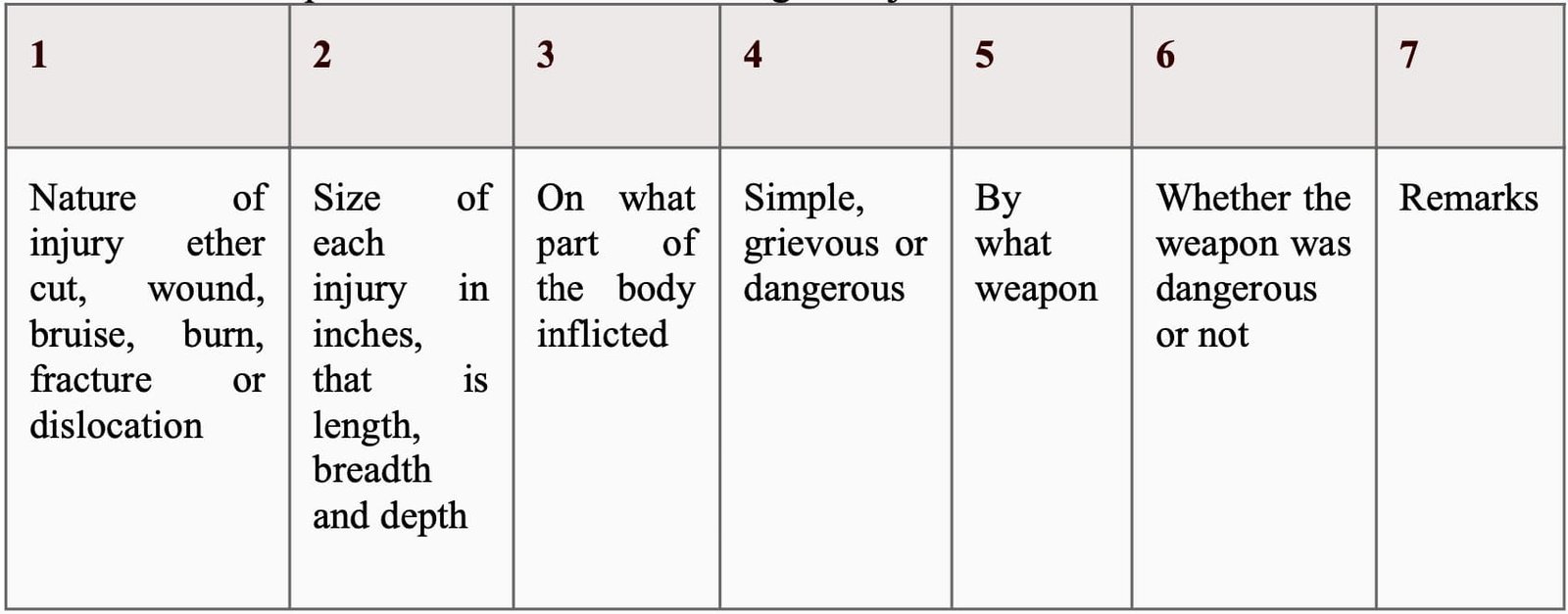
The Veterinary Officer may be supplied by the Police Superintendent or The Magistrate for veterolegal examination of the injured animal with following form which he is required to fill in after examining the injured animal.
The Veterinary Officer should be very careful in filling in this form. First of he should write at the left hand top corner of the form identification of animal and the name and number of the police constable accompanying it and should note the mark or marks of identification to enable him to recognize the injured animal. He should then note the exact time of the examination, viz. hour, date, month and year, and proceed with the examination proper as below:
Nature of Injury
While describing the injuries in columns 1, 2 and 3 of the form, the Veterinary officer should carefully note their nature and number, the character of their edges, their size as regards length, breadth and depth, the line of direction and their situation.
Presence of any extraneous material in the wound should be carefully noted. If necessary, he should use a magnifying lens. All the injuries should be measured with a tape-measure, and the exact measurements in inches must be given.
While mentioning the exact situations a reference to some bony prominences or anatomical contours should be made.
Simple, Grievous or Dangerous Injury
In column NO.4 it must be mentioned whether the injury is simple, grievous or dangerous to life. A simple or slight injury is one which is neither extensive nor serious, and which heals rapidly without leaving any permanent deformity or disfiguration.
Grievous injuries as described in Section 320 IPC, are as follows:
- Emasculation
- Permanent privation of the sight of either eye
- Permanent privation of the hearing of either ear
- Privation of any membrane or joint
- Destruction or permanent impairing of the powers of any membrane or joint
- Permanent disfiguration of the head or face
- Fracture or dislocation of a bone or tooth
- Any hurt which endangers life or which causes the sufferer to be, during a period of twenty days, in severe bodily pain, or unable to follow ordinary pursuits.
Kind of Weapon
In the fifth column the kind of weapon by which the injury was inflicted should be mentioned. In there marks column the age of injury should be noted.
Age of Injury
The age of a bruise may be ascertained from the colour changes which its ecchymosis undergoes. These changes start from eighteen to twenty- four hours after its infliction. On histological examination at necropsy the polymorphonuclear leucocytes in a haematoma begin to disintegrate after 3 to 5 hours, mostly they are fragmented within 21 hours and by 30 hours the basophilic nuclear fragments have either undergone autolysis or have been engulfed by the phagocytic monocytes.
The age of a wound may be ascertained by observing stages of its healing process. A wound, which is not thoroughly aseptic and is, gaping owing to loss of tissue, heals by the formation of granulation tissue.
The vascular endothelium shows distinct proliferative changes, and vascular buds are given off from the minute vessels at the periphery during twenty-four hours.
A complete network of new capillary vessels is formed in thirty-six to forty hours.
Spindle-shaped cells, which run at right angles to the vessels in the deeper parts of the wound, are visible in forty-eight to fifty-six hours.
Definite fibrils are seen running parallel to the long fibroblasts in three to six days.
The cellular structure and vessels are obliterated gradually, and are replaced by a dense fibrous scar tissue in three to four weeks.
Pus may appear in a septic wound in about thirty-six to forty-eight hours. In bone fracture the reparative process indicates its approximate time.
Signs of inflammation and exudation of blood in the soft parts and around the fractured ends- first to the third day.
Soft provisional callus-fourth to the fifteenth day. Ossification of callus-fifteenth day to the fifth week. Complete absorption of callus-six to eight weeks.
Bone formation does not occur in comminuted fractures. In the case of dislocation of a joint, the time can be judged from the colour changes of the bruise.
When a tooth has been hit and has dropped out, bleeding from its socket may continue for a few hours. The cavity of the socket usually fills up in seven to ten days, and the alveolar process becomes quite smooth after fourteen days.
Causes of Death from Wounds
The causes of death from wounds may be immediate or direct and remote or indirect.
Immediate or direct
Haemorrhage
Haemorrhage may be extemal or internal. External haemorrhage may produce marked fall in blood pressure and consequent shock, causing death either rapidly, if a large blood vessel, such as the carotid or femoral artery, has been wounded, or slowly, if a number of small vessels have been injured. Sudden loss of blood is more dangerous than the same quantity lost slowly. The loss of one-third of the blood in the body is almost enough to cause death. Internal haemorrhage may occur in penetrating and gunshot wounds. It need not be profuse for a fatal result; for a small quantity of haemorrhage in the brain or pericardium may prove rapidly fatal by disturbing the functions of the brain or heart owing to mechanical pressure on them. Blood flowing into the windpipe may cause death mechanically by asphyxia Rupture of internal organs like spleen, liver, lungs and heart usually causes fatal internal haemorrhage, only the symptoms are often noticed after a few hours.
Injury to a vital organ
Severe injury to a vital organ, such as crushing of the brain, heart, lungs, etc., is, as a rule, rapidly fatal.
Shock
Death may occur from shock without any visible injury due to paralysis of the heart by a hit in the cardiac region. Death from shock occurs easily in animals that are severely ill and weak, old and young.
Remote or indirect
- Inflammation of the internal organs, such as meningitis, cerebritis, pleurisy, pneumonia, peritonitis, etc.
- Septic infection of a wound causing septicaemia, pyaemia or exhaustion from prolonged suppuration.
- Gangrene or necrosis resulting from severe crushing of parts and tearing of the blood vessels or crush syndrome.
- Thrombosis in veins and embolism.
- Infective diseases, such as erysipelas and tetanus, which may develop through the entrance of the causal organism through a wound.
- Fat Embolism occurring after fracture of long bones and injury to fatty tissue.
- In injuries to the jugular, cephalic and femoral veins, air may be sucked in because of the negative pressure during inspiration and intravenous injection may cause death.
- Neglect of Injured Animal.
- Result of Surgical Operation: If death follows a surgical operation performed for the treatment of an injury caused by the offender, he shall be responsible for the result, if it is proved that the death was inevitable even without the operation and that the operation was thought necessary and was performed by a competent surgeon with reasonable care and skill.
Difference between Wounds Inflicted during Life and After Death
There is more or less copious haemorrhage in all wounds. The effused blood is forced into the tissue interspaces in the vicinity of the wounds, and is found infiltrated into the cellular and muscular tissues.
There is consequent staining of the edges of the wounds and the neighbouring tissues, which cannot be removed by washing, but the staining caused by the blood effused from post-mortem wounds is easily removed by washings.
Inhaled blood will be found in the lungs and bronchi, if the area of haemorrhage communicates with the bronchial tree. There will be clots of effused blood in the wounds and tissues, and in the neighbourhood of the body.
Clotting of the blood normally occurs in about four to ten minutes. There will also be signs of spouting of arterial blood on the body, or in its vicinity. In a contusion ecchymosis, absorption, changes in the colour and a swelling of the neighbouring tissue will be present.
On dissection coagulated blood will be found in the subcutaneous tissues. Retraction of Edges of the Wound will be present if caused in life. Likewise Signs of Inflammation and Reparative Processes will be present if made during life.

