TABLE OF CONTENTS
In Vitro Fertilization (IVF) in Animals: Step-by-Step Guide to Oocyte Retrieval, Media Preparation, and Embryo Culture
In Vitro Fertilization (IVF) is a reproductive biotechnology in which oocytes (egg cells) are collected from the ovaries of a donor animal and fertilized with sperm outside the body (in vitro), usually in a laboratory. The resulting embryos are then cultured for several days and subsequently transferred into the uterus of recipient (surrogate) cows for gestation.
In vitro fertilization (IVF) technology is also called as Ovum Pick-up and In Vitro Embryo Production (OPU-IVEP) Technology, is an advanced reproductive technology for multiplication of superior female germplasm at much faster rate.
Day 1: Media Preparation (IVM)
- Take 50 ml OCM media in a test tube and incubate 2 hours at 38.5˚C under 5 percent CO2 in air.
- Take 9 ml TCM and 1ml serum (10%) and filter using 0.2µn filter. Take 8ml of filtered media and incubate at 38.5˚C under 5 percent CO2 in air.
- In 2 ml of filtered TCMh +FBS media add 2µl FSH, 2µl LH and 2µl E2.
- Using a sterile pipette, place 50µl droplets in 35 mm petri dish and are cover with sterile mineral oil.
- Pre-equilibrate in incubator for 2 hours at 38.5˚C under 5 percent CO2 in air.
Day 2: Media Preparation (IVF)
- Take 15-20 ml sperm TALP media in a test tube and incubate 2 hours at 38.5˚C under 5 percent CO2 in air.
- Take 10 ml of IVF TALP and add 50 µl of heparin and filter using 0.2µn filter.
- Using a sterile pipette, place 75µl of IVF TALP with heparin droplets in 35 mm petri dish and are cover with sterile mineral oil and pre-equilibrate in incubator for 2 hours at 38.5˚C under 5 percent CO2 in air.
Day 3: In vitro Culture of Embryos
For Co-culture
Take 8 ml of TCMh media and add 2ml FBS and filter using 0.22µn filter. Place 50µl droplets in 35 mm petri dish are cover with sterile mineral oil and pre-equilibrate in incubator for 2 hours at 38.5˚C under 5 percent CO2 in air. Add 50 oviductal cells (explants)/droplet before embryo culture.
Day 1: In Vitro Maturation
Collection of ovaries and oocytes retrieval and maturation:
1. Ovary Collection
Materials Required
- Thermos
- Container
- 500 ml NS with antibiotic
- Forceps and scissors
- Gloves
Procedure
- Remove ovaries from the reproductive tract of animals immediately after slaughter and place the ovaries into the container containing Normal Saline (NS).
- After collection, wash the ovaries to remove blood and then transfer ovaries to the thermos containing NS at 30-35°C.
- Transport the ovaries to the lab within 2 hours of slaughter.
2. Cumulus Oocyte Complex (COCs) Collection
Materials and equipment needed for preparation COCs collection:
- BP holder
- BP blade
- Artery and thump forceps
- 90 mm integrid petri dish
- 60 mm petridish
- 50 ml test tubes
- Stereo zoom microscope
- 10 ml syringe and 18 gauge needle
- Plastic beaker
Materials and equipment needed for in vitro maturation (IVM):
- Incubator (38.5°C and 5% CO2)
- Laminar flow hood
- 10, 200, 1000 µl micropipette with sterile tips
- IVM media
- Mineral oil
- 35, 60 and 90 mm petridish
Procedure
In Laboratory, remove all adhering tissues and ligaments of the ovaries and wash thoroughly with running tap water followed by five times in normal saline supplemented with penicillin and streptomycin and one wash in 70 percent ethanol. Finally place the ovaries in NS containing beaker until oocyte collection.
In laminar flow hood, hold the ovaries with an artery forcep in a 60 mm petridish containing 3 ml of oocyte collection medium(OCM) and make cross hatched incisions on the surface of each ovary with sterile scalpel blade (No. 11) to disrupt the superficial follicles and release the COCs. Repeat the procedure upto 4-5 ovaries and transfer into 50 ml test tube. After slicing ovaries, place the test tube into an incubator for 10min. After oocytes are settled to the bottom of the tube, aspirate the sediment using micropipette and pour into the 90 mm integrid petridish containing 2 ml OCM media.
Under stereo zoom microscope, isolate the immature oocytes and transfer retrieved COCs into 35 mm petri dish containing TCM 199+10 percent FBS.
After screening under a stereo zoom microscope (Nikon, Japan) grade based on their cumulus cells investment and cytoplasm homogeneity.
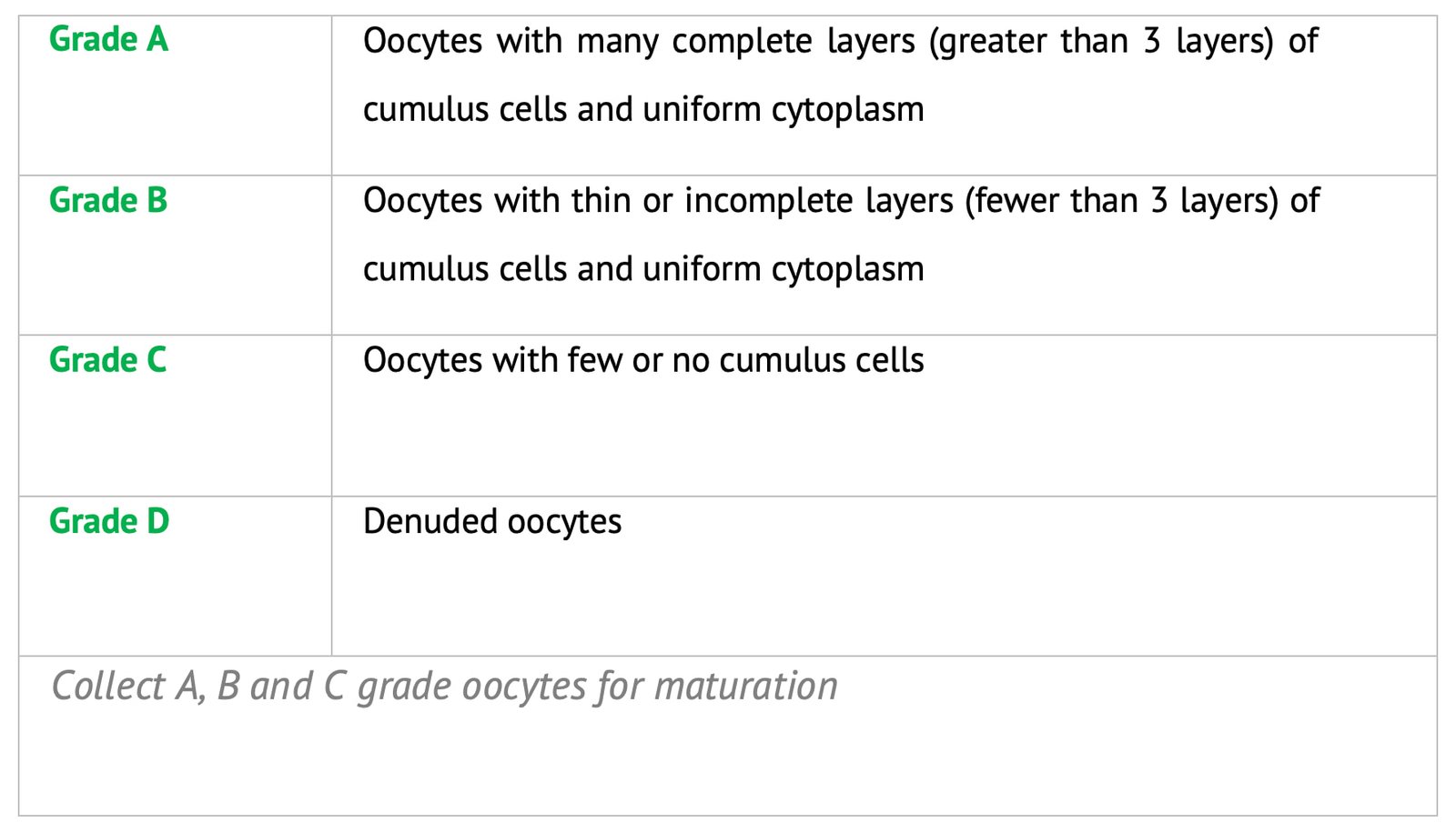
Wash the collected A, B & C grade oocytes in TCM 199+10 percent FBS three times and final wash in in vitro maturation (IVM) medium.
Transfer a group of 10 COCs to a 50 µl droplets of maturation medium in a 35 mm petri dish and incubate for 24 hours for sheep and 27 hours for goat.
Day 2: In Vitro Fertilization (IVF)
Sperm Preparation
Materials and Equipment
- Laminar flow
- Incubator
- Stereo zoom microscope
- Phase contrast microscope
- 35 mm petri dish
- Micro pipettes (10, 200 and 1000 µl)
- 50 ml and 15 ml test tubes
- Sperm TALP and IVF TALP media.
Procedure
- Collect the animal testis from the slaughterhouse and transport to the Assisted Reproductive Technology Laboratory in 0.9 percent normal saline containing penicillin (100 IU/ml) and streptomycin (50 mg/ml) at 30-35°C within 2 hours of slaughter.
- Remove the adherent tissues of the testis and wash thoroughly with running tap water followed by with 0.9 percent normal saline.
- Remove the tunica albuginea by small incision and locate the cauda epididymis.
- Locate the area below the deferent duct and make incision by sterile blade and scoop the secretions and mix with the 2 ml of pre-equilibrated Sperm-Tyrode Albumin Lactate and Pyruvate (SpTALP) medium in 35 mm petri dish and analyse the initial motility.
- Dilute 2 ml of semen with 5 ml of pre-equilibrated SpTALP by centrifugation at 1200 rpm for 5 min at room temperature (RT). Remove the supernatant and then add fresh SpTALP and repeat the above procedure for two times.
- Add 1ml of SpTALP in medium in three sugar tubes. Finally add 200 µl of the sperm pellet under 1 ml of SpTALP medium in each tube and incubate at 45° angle for swim up at 38.5°C in 5 percent CO2 in air for 45 min.
- At the end of incubation take the superficial layer of 0.4-0.6 ml of the medium containing the motile fraction from each tube and pool in a 15 ml centrifuge tube, and wash it with 10 ml SpTALP by centrifugation at 1200rpm for 5 min at RT.
- After centrifugation, remove the supernatant and collect sperm pellet.
- Determine the concentration of the final sperm pellet with a haemocytometer and dilute the sample with IVF TALP to yield a concentration of 1-2×10⁶ sperm/ml. For this, add 10 µl sperm suspension to 90 µl water to kill sperm. Load 10 µl of sample onto a hemocytometer. Count the sperm in 5 squares and multiply sperm number by 5,00,000 to determine concentration per ml. Dilute the sperm using IVF TALP.
- Oocytes preparation for Fertilization: At the end of maturation period, remove the dish from the incubator and examine under stereo zoom microscope.
Morphological Assessment of Oocytes for in Vitro Maturation
Assess the maturation rate based on the degree of cumulus expansion.
| Degree 2 | Cumulus cells were homogenously spread and clustered cells were no longer present, (Full cumulus cell expansion). |
| Degree 1 | Cumulus cells were slightly expanded and clustered cells were still observed, (Moderate cumulus cell expansion). |
| Degree 0 | No morphological change compared with fresh COCs, (Slight or no expansion). |
The 2 and 1 degree of oocytes with cumulus expansion are considered as matured. Wash the matured oocytes with TCM 199 supplemented with 500 IU/ml hyaluronidase, for 30 sec followed by gentle pipetting for partial removal of the cumulus cells in sperm-TALP media followed by two washes in IVF-TALP and then transfer to the pre-equilibrated IVF droplets (10-15 oocytes each) and incubate until fertilization.
Fertilization
Inseminate the motile sperm suspension obtained by swim up technique to the IVF droplets containing oocytes to achieve the final concentration of 2 million sperm/ml and co-incubate for 24 hours at 38.5°C in 5 percent CO2 in air.
Day 3: Culture of Presumptive Zygotes in Embryo Culture Media (18-22 hours post IVF)
Materials Required
- Laminar air flow
- Stereo zoom microscope with stage warmer
- Microcentrifuge tubes (1.5 ml)
- Vortexer
- Micropipette with sterile tips IVC media and droplets
- 35 mm petridish
- 15 ml centrifuge tubes
Procedure
- Remove the fertilization dish from the incubator and transfer all presumptive zygotes into micro centrifuge tubes.
- Remove cumulus cells and sperms from embryos by vortex the tube containing presumptive zygotes or gentle pipetting using hyaluronidase.
- Wash the presumptive zygotes three times in IVC medium and transferred into pre- equilibrated 50 µl IVC droplets (10-15/droplet). In co-culture systems add 50-100 BOEC cylinders to the droplets before culture of the IVF embryos.
Embryo Culture
- Day 2 and 5 of culture remove 10 µl of culture media and add 10 µl of fresh IVC medium to each drop.
- Assess the cleavage rate at 2nd day of culture by number of embryos cleaved divided by the number of fertilized oocytes cultures initially in the droplets. Assess embryo development in every 24 hours post insemination up to 7 days. Record the number of embryos at various stages viz. 2-, 4-, 8-16 cells, morula and blastocyst.
Embryo Quality Analysis
- Fix the different stage of embryos in 2.5 percent gluteraldehyde in DPBS at room temperature for 15 min.
- After that wash embryos in PBS/PVP three times and stain the embryos in a minimal volume of 0.1 mg/ml of DAPI for 15 min.
- After staining wash the embryos twice in PBS/PVP to remove stain particles, mount on poly-1-lysine (Sigma cote-Sigma) slides and cover with a coverslip supported with vaseline paraffin wax (9:1 w/w).
- Evaluate the nuclear morphology of embryos under fluorescent microscope with a UV filter. Nuclei appeared blue in colour. Count the no of nuclei in each embryo.

