TABLE OF CONTENTS
Classification of Placenta Based on Separation Between Fetal and Maternal Blood Supply
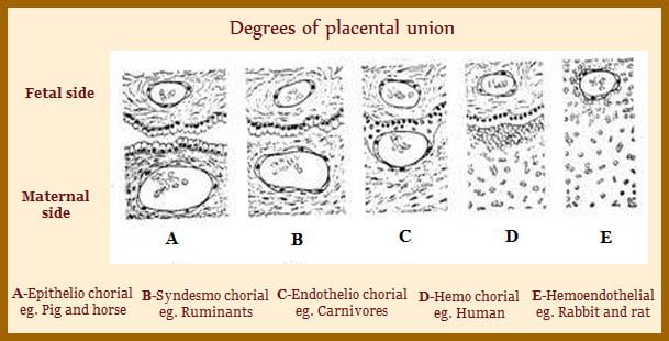
Classification of placenta based on separation between fetal and maternal blood supply is epitheliochorial, endothelial chorial and hemochorial.
The nomenclature for describing placental intimacy is derived by describing the tissues of the maternal placenta in prefix of the word.
The tissues of the fetal placenta constitute the suffix. Exchange can occur through as many as six layers and as few as three.
- Prefix: maternal side Epithelio
- Suffix: fetal side Chorial
Epitheliochorial Placenta
In this type, both the endometrial epithelium (maternal side) and epithelium of the chorionic villi are intact. There is complete intact layer of epithelium in both maternal and fetal components. Found in sow and mare;
Ruminants are also having epitheliochorial placenta. However the endometrial epithelium transiently erodes and then regrows causing intermittent exposure of maternal capillaries to the chorionic epithelium and it has been termed as syndesmochorial.
Pig, Horses and Ruminants
- Chorionic capillaries
- Chorionic interstitium
- Chorionic epithelium
- Endometrial epithelium
- Endometrial interstitium
- Endometrial capillaries
In addition to feature of partial erosion of the endometrial epithelium, a unique cell type is found in the ruminant placenta. These cells are called nucleate giant cells. They are characterised by quite large and have two nuclei. Binucleate giant cells appear at about day 14 in sheep and between day 18 and 20 in the cow.
Binucleate giant cells (BNGC) migrate from the chorion to the endometrial epithelium in ruminants. These cells are thought to secrete placental lactogen and pregnancy specific protein B (PSPB).
These cells originate from trophoblast cells and are believed to be formed continuously throughout gestation. Binucleate giant cells constitute around 20 % of the fetal placenta. During development, the binucleate giant cells migrate from the chorionic epithelium and invade the endometrial epithelium. These cells are believed to transfer complex molecules from the fetal to maternal placenta. There is evidence that they secrete placental lactogen and pregnancy specific protein B (PSPB) that are called pregnancy associated glycoproteins. These proteins are unique to pregnancy in ruminants. These binucleate cells are important sites for steroidogenesis producing progesterone and estrogen.
Endothelial Chorial Placenta
It is characterised by having complete erosion of the endometrial epithelium and underlying interstitium. Thus maternal capillaries are exposed to epithelial cells of the chorion.
Dogs and Cats
- Chorionic capillaries
- Chorionic interstitium
- Chorionic epithelium
- Endometrial interstitium
- Endometrial capillaries
Haemochorial Placenta
The haemochorial palcenta is characterised by having chorionic epithelium in direct apposisition to maternal pools of blood.
Thus, nutrients and gases are exchanged directly from maternal blood and must move through only tissue layers. This highly intimate relationship is found in primates and rodents.
- Chorionic capillaries
- Chorionic interstitium
- Chorionic epithelium

