TABLE OF CONTENTS
Cataract in animals
Cataract is the most common surgical affection of lens of eye in animals. Opacity of the lens is known as cataract. It is a degenerative lesion of the lens.
Cataract in a cow
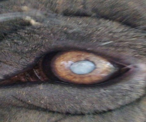
Cataract in a lion
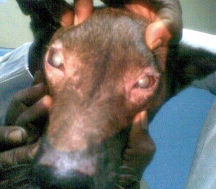
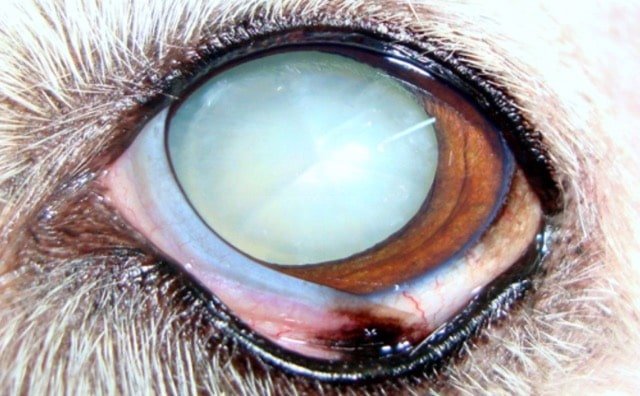
Bilateral Cataract in a dog
Cataract in a Elephant
Classification of Cataract
Congenital cataract
Congenital cataract is the cataract present at birth. (In foetal life the lens receives its nutrition through vascular channels. After birth the lens is entirely dependent on the aqueous humour for its nutrition. In puppies and kitten it is normal for the vascular covering of the foetal lens to persist for a few days after the eyes have opened. This should not be mistaken for congenital cataract.)
Acquired cataract
Acquired cataract is cataract that developing later in life.
Cataract may be complete (involving the lens completely), partial, progressive or stationary.
Juvenile cataract
Juvenile cataract is cataract that seen in young animals.
Senile cataract
Senile cataract is cataract that developing due to old age. This is common in veterinary practice.
Diabetic cataract
Diabetic cataract also is not seen in veterinary practice. Diabetic cataract is characterized by minute opacities developing on the superficial cortex of the lens due to turgidity of cells in the superficial cortex of the lens. The turgidity of cells is apparently associated with the sugar content of aqueous humour.
Toxic cataract
Toxic cataract is cataract that caused by the circulation of toxins or poisons in the body, e.g., cataract due to equine influenza, periodic ophthalmia, distemper, chronic nephritis, ergot poisoning in cattle and pigs, experimental feeding of naphthalene, etc.
Cortical cataract
Cortical cataract (Anterior cortical and posterior cortical cataracts) majority of cortical cataracts are stellate cataracts, i.e., spreading from the centre of the lens to its periphery. Cortical cataract sometimes develops as a complication of a perforating corneal ulcer.
Pyramidal cataract
Pyramidal cataract is a localized opacity of the lens.
Lamellar cataract
Lamellar cataract is the opacity is seen in the area between the lens nucleus and cortex.
Perinuclear cataract
Perinuclear cataract is lamellar cataract seen in horses.
Nuclear cataract
Nuclear cataract is confined to the central portion (nucleus) of the lens.
Diffuse cataract
Diffuse cataract is spreading evenly through the entire lens substance.
Calcareous cataract
Calcareous cataract is a cataract in which the lens substance is partly converted into chalky materials.
Depending on the stage for surgical removal of the lens cataract is describes as-
In hyper-mature cataract there may be partial calcification of the lens and some portion of the lens may also undergo liquefaction. The cortex appears milky white in colour. The nucleus of the lens may sink into the bottom of liquefied lens substance. Complete removal of the lens is difficult when the cataract is hyper – mature.
Etiology
- There might be a hereditary predisposition.
- Toxins
- Senile changes attended with old age.
- As a sequelae of diseases of the eye like irirdocyclitis or systematic diseases like diabetes.
Prognosis
For juvenile cataract seen in young animals, the prognosis is good.
Diagnosis
The pupil is dilated by instilling tropicamide (2%) into the eye, in order to facilitate examination of the lens.
The diagnosis of Cataract in animals can be made by using an ophthalmoscope.
Treatment
Discission or Needling
The anterior capsule of the lens is incised in a cruciate fashion, using a cataract needle so that the aqueous humour will come in contact with the lens substance and will facilitate re-absorption of the opacity.
Discission will have to be repeated periodically to obtain the desired effect. It may not be effective in all cases.
Couching of the lens
The lens is pushed downwards and backwards by introducing a proper instrument through an incision in the cornea. Couching of the lens is not a practicable treatment in veterinary practice because the suspensory ligament of the lens in animals is very strong.
In man, dissolution of the opaque lens by using 0.02% trypsine has been reported.
Removal of the lens
Removal of the lens is occasionally done in the dog. The removal of the lens in the dog is more difficult than in man because the lens of the dog is proportionately much larger, and the suspensory ligament of the lens is tough.
Removal of the lens will not serve any purpose if there are degenerative changes in the retina associated with cataract. In estimating the prospects of the operation the existence of pupiliary reflex is of some help.
There are two methods for removal of the lens, viz., the intracapsular extraction, and the extracapsular extraction.
Intracapsular extraction is the extraction of the lens with its capsule. This is difficult in animals because of the tough suspensory ligament.
Extracapsular extraction is the extraction of the lens without its capsule. The anterior capsule of the lens is incised and through that the lens substance is removed.

Extracapsular extraction is successful only if the cataract is ripe (mature). At this stage the endothelial cells of the capsule that are left behind are incapable of proliferating. Whereas, if the operation is done before fully ripe, the proliferation of the endothelial cells after surgery may once again create opacity and this will interfere with vision.
Extracapsular extraction of the lens is difficult if the cataract is hyper – mature because of the partial liquefaction or softening of the lens substance.
ECCE +IOL ( Extra capsular catarct extraction + Intra ocular lens)
Extracapsular cataract extraction is an operation in which the lens nucleus and cortex, excluding the capsule, are delivered through a corneal incision involving about one-half of the circumferences of the cornea, with rigid lens implantation.
Technique of Removal of Lens
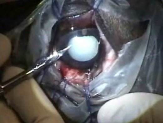
The operation is done under general anesthesia. The eyeball is fixed during the operation by one or more stay sutures passed through the sclera.

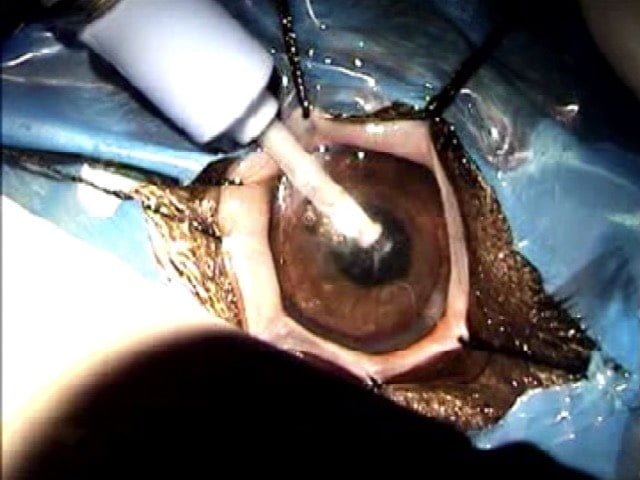
Eye ball positioned for cataract surgery in a dog
The cornea is punctured above the 3 – O – clock point, about 0.1 cm away from the limbus. By using a small scissors or the Graefes’ knife the incision is extended upwards along the cornea parallel to the limbus, to the 9 – O – clock point.
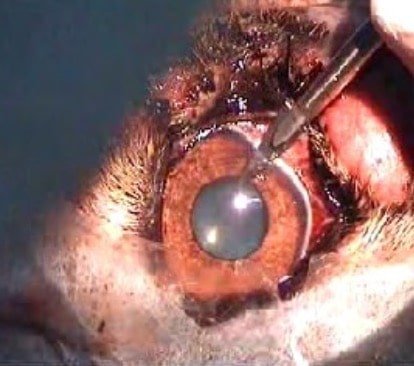
In intracapsular extraction, the lens is held close to its equator with a special forceps (Duthies’ forceps or Arrugas’ forceps) and then it is gently moved and detached fromm its suspensory ligament.
If extracapsular ectraction is desired,the anterior capsule of the lens is incised in the form of a T and a cataract scoop is used to remove the lens substance.
After removal of the lens the corneal wound is sutured. Conjunctival keratoplasty is advisable. Postoperative interference by patient should be guarded against. For this it is desirable to administer sedatives for at least two or three days.
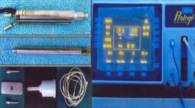
Phacoemulsification and IOL implantation.
Phacoemulsification is a procedure in which the lens is ultrasonically fragmented and aspirated through an incision about 3.2 mm and extended upto 8mm and 4.2mm with rigid and foldable lens implantation respectively.
Phacoemulsification in progress