TABLE OF CONTENTS
Blood Testis Barrier (BTB) in Animals
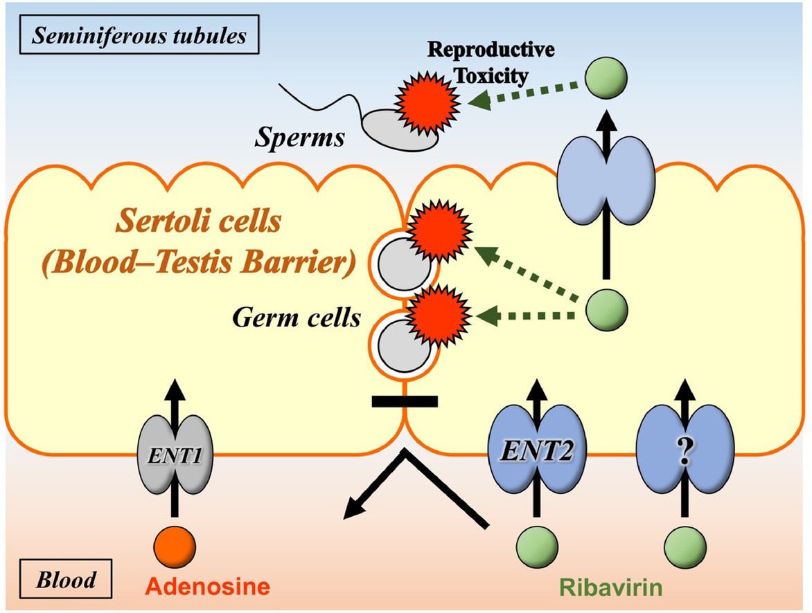
The blood testis barrier (BTB) in animals is formed by tight junctions between adjacent Sertoli cells within the seminiferous tubules to create a specialized environment that protects developing germ cells from the immune system, maintains compartmentalization for spermatogenesis, and regulates the movement of molecules between the blood and seminiferous tubules.
Due to the blood testis barrier, the seminiferous tubules are not penetrated by blood and lymph vessels. The chemical changes that occur in the blood cannot occur within the seminiferous tubules.
The peritubular cells surrounding the seminiferous tubules and the Sertoli cell junctional complexes form the blood testis barrier.
Its primary function is to prevent autoimmune reactions from destroying the developing germ cells.
Myoid Cells
Myoid cells are the incomplete or partial barriers located in the basement membrane of seminiferous tubules. This barrier is poorly developed in bull, ram and boar. It is not important in farm animals.
Sertoli Cell Junction
The tight junctions formed between two adjacent sertoli cells divide the germ cells in two compartments as basal compartments and adluminal compartments. They are the true blood testis barrier.
Importance of Blood Testis Barrier
- Prevents autoimmune destruction of developing germ cells
- Divides seminiferous tubules into basal and adluminal compartments
- Maintains a controlled environment for spermatogenesis
- Limits the entry of toxins and pathogens
- Ensures selective transport of molecules
- Supports proper germ cell maturation
- Provides structural integrity to seminiferous tubules