TABLE OF CONTENTS
Testis (Testes) in Animals: Development, Features and Structure
The testis is the primary andrological organ responsible for producing spermatozoa and the male sex hormone, testosterone. Morphologically, it is an oval-shaped, paired gland. The right and left testicles are separated by a muscular septum formed by the dartos muscle.
The testis, also referred to as the orchium or male gonad in male animals, derives its name from the Greek word ‘orchis’.
“Testis” and “testes” refer to the same organ, but differ in number:
- Testis: singular
- Testes: plural
- Testicles: Casual term and often used in non-scientific discussions
It is located outside the abdominal cavity. In most species, the testes are situated in the inguinal region within the scrotum; however, in elephants, whales, seals, dolphins, birds, and rhinoceroses, they remain intra-abdominal.
Each testis is supported in one of the two scrotal pouches, where it is held in place by its tunics and the spermatic cord.
The spermatic cord is composed of the following structures:
- Spermatic artery
- Spermatic vein
- Spermatic nerve
- Internal cremaster muscle
- Lymphatic vessels
- Vas deferens
- Tunica vaginalis propria
Embryonic Development
The origin of the testes is initiated from the gonadal (genital) ridge and occurs in four phases:
- First Phase: Migration of germ cells to the gonadal ridge.
- Second Phase: Formation and proliferation of the blastema, leading to the development of the indifferent gonad.
- Third Phase: Migration of mesonephric cells into the gonad.
- Fourth Phase: Sexual differentiation and development into the testis.
Structure
The testis consists of the following main components:
- Testicular capsule
- Parenchyma
- Mediastinum testis
- Rete tubules (Rete testis)
The testicular parenchyma consists of the following structures:
- Seminiferous tubules
- Interstitial cells of leydig
- Capillaries
- Lymphatic vessels
The testis proper is covered by two capsules:
- Tunica vaginalis propria
- Tunica albuginea
Tunica vaginalis propria is composed of:
- Parietal layer: Faces the scrotum
- Visceral layer: Faces the testis
Internal Features
The distal end of the testis is attached to the scrotum by the scrotal ligament.
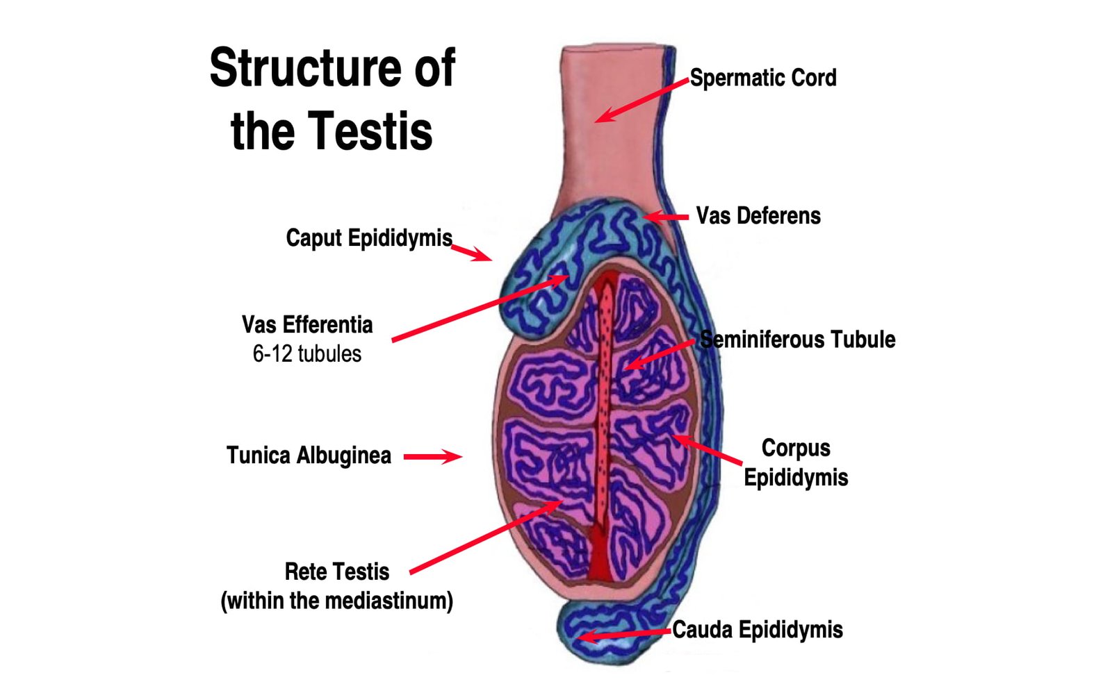
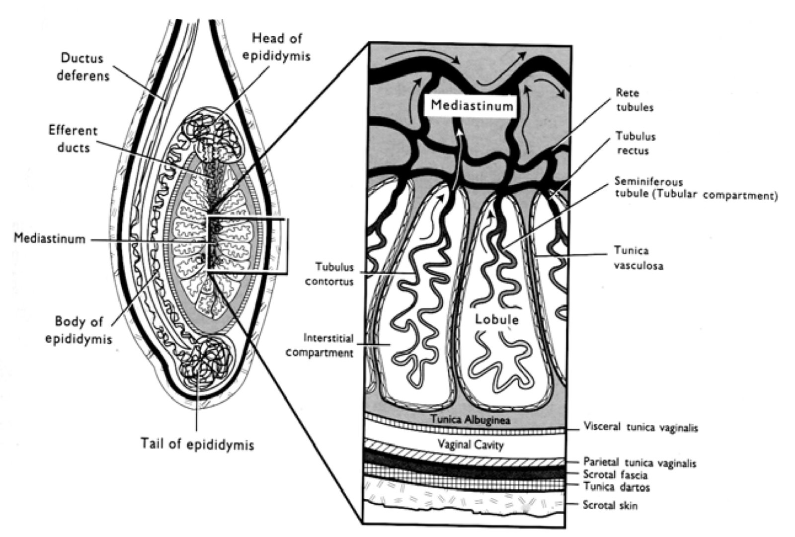
The testis proper is closely covered by a thin serous membrane called the tunica vaginalis propria. Beneath this structure is a dense, thick connective capsule, the tunica albuginea, from which septa radiate toward the mediastinum testis, except in the stallion, where it forms the lobules of the testis.
Within these lobules are the seminiferous tubules, which are lined by germinal epithelium that produces spermatozoa, and Sertoli cells, which are located between the developing germ cells.
The Sertoli cells are also known as nurse cells, pillar-like cells, or sustentacular cells.
The interstitial cells (Leydig), located between the seminiferous tubules, secrete male hormones.
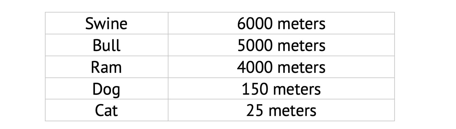
Approximate length of seminiferous tubule:
The cross-section of seminiferous tubules contains three layers:
- Outer capsule
- Basement membrane
- Testicular cells
Testicular cells are composed of:
- Germinal cells
- Parenchymal cells or testicular somatic cells
- Sertoli cells
The seminiferous tubules converge at the apex of the lobule at the receptacle, joining the straight tubules.
The straight tubules, also called tubuli recti, enter the rete testis, a structure of anastomosing spaces located in the mediastinum testis.
The mediastinum testis is absent in the stallion, unlike in other animals. The collecting tubules join the efferent tubules.
From the rete testis, 6 to 24 efferent ducts arise. These ducts then form a single duct called the caput epididymis.
Upon examination of the testes, the tortuous configuration of blood vessels is most readily noted in the tunica albuginea in the bull. This configuration plays a role in assisting the heat regulatory mechanism of the testis.
The consistency of the testis is typically turgid. The parenchyma is yellow to reddish-brown in color and bulges upon sectioning.
Physiological Functions
- It produces male sex hormone(androgen) and inhibin
- It secretes testicular fluid
- It nourishes the newly born male gamete
- It helps in the transportation of spermatozoa
- It secretes estrogen in limited amount
Associated Structures
- Testicular ligament
- Testicular mesentery
- Testicular appendix
Testicular Ligament
Fetal ligament which is a derivative of gubernaculum and present during the descent of the testis in to the scrotum. Later on, it gets atrophied.
Testicular Mesentery
The testicular mesentery is a part of the primitive mesentery that encloses the fetal testis and is present during its descent into the scrotum. In postnatal life of livestock, it continues as a peritoneal fold between the testis and the epididymis.
Testicular Appendix
Testicular appendix is non-functional residual part of embryonal hood during post-natal life of livestock.
Blood Supply
The testis is richly supplied with blood by the Spermatic artery/Testicular artery, a branch of the abdominal aorta.
The veins on leaving the testicles form a network, the pampiniform plexus around the artery in spermatic cord. The spermatic vein which issues from this plexus, usually joins with the posterior venacava on the right side, the left renal vein on left side.
Nerve Supply
The nerves derived from the renal and posterior mesenteric plexus form the spermatic plexus around the vessels to which they are chiefly distributed.

