TABLE OF CONTENTS
Surgical affections of soft and hard palate
Surgical affections of soft and hard palate in animals are cleft palate or palatoschisis, aquired oronasal fistula, lampas or palatitis or lampers, Neoplasia, protrusion of soft palate etc.
Surgical affections of soft and hard palate listed-
- Cleft palate or Palatoschisis
- Aquired Oronasal fistula
- Lampas or Palatitis or Lampers
- Neoplasia of palate
- Protrusion of soft palate
Cleft palate or Palatoschisis
Cleft palate or Palatoschisis or Congenital Oronasal Fistula is an abnormal communication between the oral and nasal cavities involving the following structures- soft palate, hard palate, premaxilla and with or without lips cleft.
The Lip and Premaxilla contributes to the primary palate, and incomplete closure of this is a primary cleft or cleft lip (harelip).
The hard and soft palates contribute to the secondary palate, and incomplete closure of either of these structures is called a secondary cleft or cleft palate.
Clinical signs of Cleft palate
The cleft is present at birth. Not always recognized immediately. Brachycephalic breeds are more commonly affected. In cats Siamese breed is usually affected.
Nasal regurgitation of milk during or after nursing, respiratory infection and failure to thrive are the major problems associated with this condition.
There will be incomplete closure of the lips, incomplete closure of the premaxilla, hard palate, or soft palate.
Treatment of Cleft palate
Tube feed the animal (via esophagostomy or gastrostomy tube) to maintain an adequate nutritional status and to reduce the incidence of aspiration pneumonia until they are old enough for surgery.
Surgery is performed when the animal is above 2 months of age, because the puppies will be better able to metabolise the anaesthetic drugs and hence lesser anaesthetic risks.
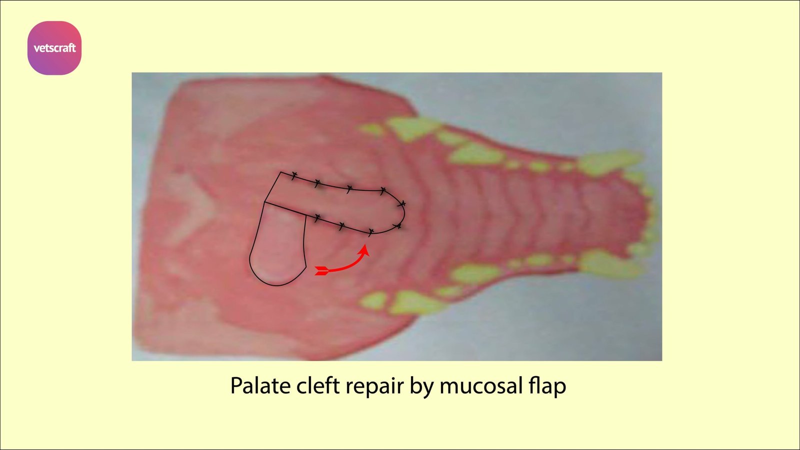
Techniques of closure of Hard palate defects by surgery-
- Sliding bipedicle flap technique
- Overlapping flap technique
Sliding bipedicle flap technique
In Sliding bipedicle flap technique, mucoperiosteal incisions are made on either side of the cleft and the mucoperiosteum is elevated from the hard palate with the major palatine artery.
The nasal mucosa and mucoperiosteum are then apposed in two layers over the defect in the hard palate.
Overlapping flap technique
Mucoperiosteal flap is made on one side of the cleft, and rotated medially to cover the hard palate defect.
The edge of this flap is inserted between the hard palate and the mucoperiosteum on the opposite side of the defect.
The flap is secured in position with horizontal mattress sutures. Lateral relief incisions are made to reduce tension on the repair.
Closure of Primary clefts involving the lip, pre-maxilla and nostril-
A mucosal flap is created from the nasal wall and sutured to a labial mucosal flap to separate the nasal cavity from the oral cavity. The cleft lip is then repaired with one or a series of Z plasties.
Complications and Prognosis of Cleft palate
Dehiscence and incomplete healing are the most common complications.
Dehiscence of hard palate repair occurs due to excessive tension and motion of the tongue against the repair.
In case of repair of the lip, dehiscence occurs if the orbicularis oris muscle has not been apposed; which causes excess tension on the suture line during movement of the lip.
Late dehiscence occurs due to growth-induced stress on the repair and can be treated when the patient matures. Prognosis is good; however several operations may be required.
Acquired Oronasal fistula
Acquired Oronasal fistula is the abnormal communication between the nasal and oral cavities caused by trauma or disease.
Acquired Oronasal fistula is most commonly caused by dental disease- when a deep maxillary periodontal pocket progresses to the apex of the tooth, lysing the bone between the apex of the alveolus and the nasal cavity or maxillary sinus.
Foreign bodies lodged between the dental arcades may cause pressure necrosis of the hard palate and subsequent development of an oronasal fistula.
Clinical signs of Acquired Oronasal fistula
Any breed or gender may be affected. Oronasal fistula occurring secondary to dental disease or tumous are seen more often in middle-aged and older animals.
That developing secondary to trauma may occur at any age. Ingested food that passes through the fistula into the nasal cavity may be expelled from the nostril by sneezing.
Chronic rhinitis is common in Acquired Oronasal fistula.
Treatment of Acquired Oronasal fistula
Treatment of Acquired Oronasal fistula is surgery. these techniques followed-
- Direct apposition
- Single-layer flap repair
- Double-layer flap repair
Direct apposition
Direct apposition of the fistula is performed only if the fistula is very small. The mucosa around the fistula is incised.
The gingival flaps are elevated and the edges of the fistula are debrided. The mucosa is then apposed over the defect.
Single-layer flap repair
If the fistula is between the gingival and buccal mucosa, the fistula is debrided and a buccal flap is advanced over the defect.
A rotational flap is done by debriding the fistula and rotating a mucoperiosteal hard palate flap over the defect.
To repair lesions at the junction of the hard and soft palates, debride and close the defect with a soft palate advancement flap.
Double-layer flap repair
This may be performed using tissue surrounding the fistula and a flap from the mucoperiosteum of the hard palate.
Create the first flap by rotating the gingival margins of the fistula medially and apposing with sutures. This flap is covered with a rotational mucoperiosteal hard palate flap.
Lampas or Palatitis or Lampers
Lampas or Palatitis or Lampers is the inflammatory ridge like thickening of mucous membrane of hard palate immediately posterior to the upper incisors in equines. Common in young horses during the development of teeth.
Treatment of Palatitis
Anti-inflammatory drugs, soft food should be advised.
Neoplasia of palate
Epistaxis is common symptom in Neoplasia of palate in animals.
Squamous cell carcinoma of nasal maxillary sinus, Osteogenic sarcoma are common tumors of palate with facial bones involving the palate.
Protrusion of soft palate
Soft palate of male camel is distended by expiratory air and protrudes out of the oral cavity while creating oral sounds. Injuries by molars or canines or violence that leads to bleeding and submucosal haematoma, ultimately this leads necrosis and gangrene.
Treatment of Protrusion of soft palate
Amputation of the affected part is done to treat protrusion of soft palate.