TABLE OF CONTENTS
Surgical affection of liver
Surgical affection of liver are Portosystemic shunts (Portosystemic vascular anomalies), Neoplasia of liver, Hepatic abscess, Liver trauma, Cholelithiasis etc.
Surgical affection of liver in animals are-
- Portosystemic shunts (Portosystemic vascular anomalies)
- Neoplasia of liver
- Hepatic abscess
- Liver trauma
- Cholelithiasis
Portosystemic shunts (Portosystemic vascular anomalies)
Blood draining from the stomach, intestines, pancreas and spleen (portal blood) has to pass through the liver before going into the systemic circulation.
Portosystemic shunts are abnormal vessels through which the portal blood bypass the liver and enter the systemic circulation.
Two main types of Portosystemic shunts-
- Extrahepatic– shunts located outside the liver parenchyma
- Intrahepatic– shunts located inside the liver parenchyma
Portocaval shunt is portosystemic shunt from portal vein to caudal vena cava.
Diagnosis
Diagnosis is from the signalment (usually purebred dogs are at increased risk), from history (failure to grow, small body stature or loss of body weight and varied signs), from physical examination findings (like microhepatica, prominent kidneys, neurological abnormalities)
Confirmation of diagnosis
- Contrast Radiography
- Ultrasonography and
- Nuclear Imaging
Treatment
Surgical correction which aims at attenuation of the shunts in case of portosystemic shunts.
Neoplasia of liver
Primary and Metastatic tumors usually seen in liver of animals.

Primary tumors
- Less common compared to Metastatic tumors
- Hepatocellular carcinomas. May develop as solitary mass or in diffuse multiple nodules
- Bile duct carcinomas
- Hepatomas
Metastatic tumors
- More common than primary tumors
- Originating from the spleen (haemangiosarcoma)
- Originating from the colon (adenocarcinoma)
- Originating from the pancreas (adenocarcinoma, islet cell carcinoma)
- Originating from the lymph nodes (lymphosarcoma)
Clinical signs
- Weight loss
- Cachexia
- Jaundice
- Ascites
- Anaemia
- Vomiting
- Diarrhoea
Treatment
Radical surgery involves excision of the mass by-
- Wedge resection
- Finger fracture technique
Large masses can also be treated by total or subtotal lobectomy.
Hepatic abscess
Hepatic abscess rare in dogs and cats but may be the result of infection. May develop due to haematogenous spread of infectious agents, penetrating foreign objects, extension of biliary infections, or localized peritonitis (necrotising pancreatitis).
Symptoms
- Prolonged and undulant fever (Suspect hepatic abscessation in case of pyrexia of unknown origin)
- Anorexia
- Abdominal pain
- Vomiting
Diagnosis
- Serum biochemistry
- Ultrasonography
- Radiography
Treatment
- Surgical drainage (Preferable for solitary abscess)
- Antimicrobial therapy
Liver trauma
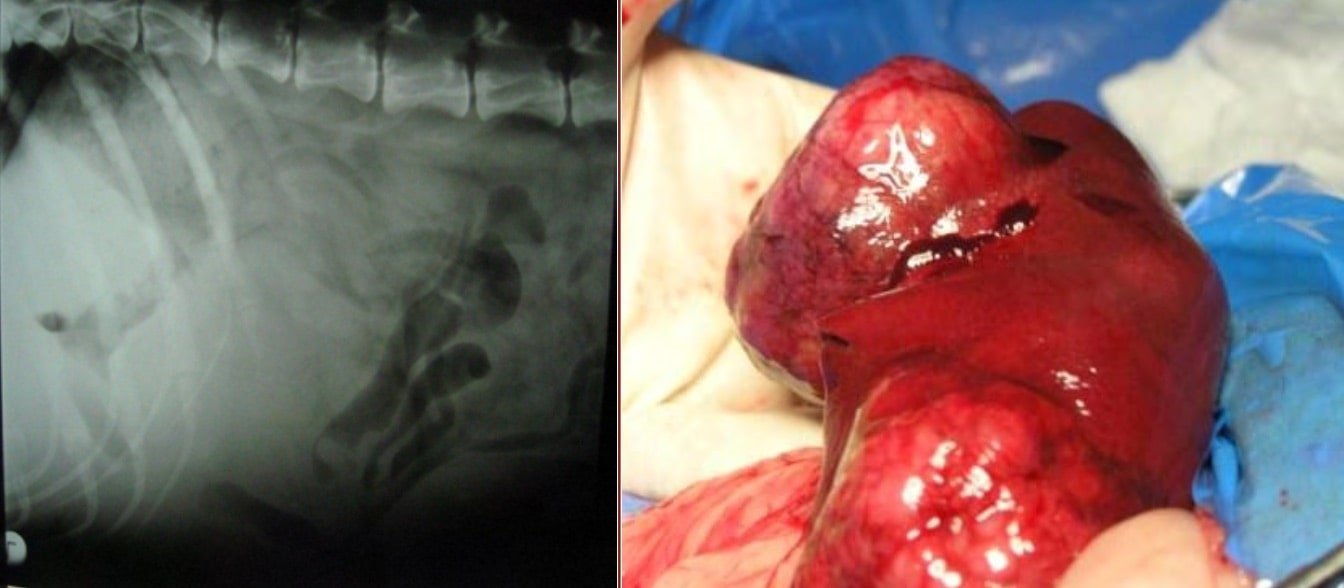
Liver trauma may be due to automobile accidents, gunshot wounds, falling from heights and rupture of necrotic tumors.
Liver trauma can be classified as-
- Transcapsular trauma
- Subscapular trauma
- Central trauma
Symptoms
- Hypovolaemia due to Acute blood loss
- Endotoxaemia (coliform or anaerobic)
- Bile peritonitis
Diagnosis
- From Haematology and Serum biochemistry
- Radiography and Ultrasonography
- From peritoneal lavage and centesis
Treatment
- Management of shock
- Control of haemorrhage
- Surgery should aim at excision of dead liver tissue, suturing the lacerated liver tissue, control of haemorrhage, and drainage of bile contents from the peritoneal cavity
Cholelithiasis
Cholelithiasis also known as gall stones and is rare in dogs.
Cholelithiasis causes obstruction to the flow of bile. The stones are formed by the precipitation of supersaturated cholesterol or bilirubin in the bile.
Obstruction to the flow of bile leads to subsequent clinical signs.
Symptoms
- Abdominal pain
- Vomiting
- Steatorrhoea and jaundice
Diagnosis
- Contrast Radiography (Intravenous or oral cholecystography) because majority of the gall stones may be radiolucent
- Ultrasonography
Treatment
Incision of common bile duct and removal of the gall stones, Cholecystotomy.