TABLE OF CONTENTS
Sperm Abnormalities: Classification, Examination Methods and Eosin-Nigrosin Staining
Sperm abnormalities refer to any deviations from the normal structure, shape, or morphology of spermatozoa.
Every ejaculate will have some abnormal sperms in it. An increased prevalence of abnormal sperm is associated with a decrease in fertility.
The appearance of an increased number of abnormal sperm in the ejaculate is a reflection of lesions of the testes and/or the excurrent duct system and provides a convenient clinical diagnostic aid.
Classification of Sperm Abnormality
Any deviation from normal morphological structure of spermatozoa is called as abnormal sperm.
There are different types of classifications available. They are:
- Based on source of abnormality
- Based on effect on fertility
- Based on the location of abnormality
1. Based on Source of Abnormality
Primary Abnormality
- They are appearing at the time of formation of spermatozoa.
- This is mainly caused due to aberration in spermatogenesis.
- These abnormalities are arising due to developmental defects in seminiferous tubules of diseased, degenerative or hypo plastic or inherited sterile conditions.
Secondary Abnormality
These abnormalities are acquired at the time of transport of spermatozoa through epididymis, accessory sex glands and urethra.
Tertiary abnormality
Tertiary abnormality caused by the semen handler after collection. It is mainly due mishandling.
Primary abnormalities are dangerous and they can be transmitted to their young ones also.
2. Based on the Effect on Fertility
- Major abnormalities: Abnormalities which are having more effect on fertility is called as major abnormalities.
- Minor abnormalities: Some abnormalities will have lesser effect and is called as minor abnormalities.
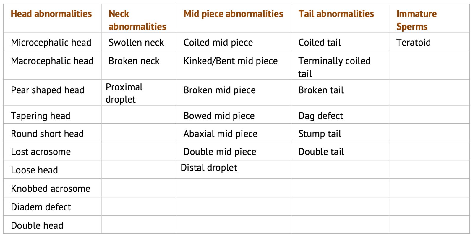
3. Based on the Location of the Abnormality
This depends upon the region where the abnormality is located. It is divided in to head, mid piece and tail abnormalities.
Methods of Assessing Sperm Morphology
Sperm morphology is assessed by:
- Eosin-Nigrosin stain
- Rose Bengal stain
- Indian ink stain
- Wright’s stain
- Casaretts’s stain
- Buffered formal saline method (wet smear method)
An eosin-nigrosin stain is commonly used as a morphology stain. The eosin-nigrosin sperm smears prepared for live and dead count is satisfactory for assessing abnormal spermatozoa.
Eosin-Nigrosin Staining Method
Materials Required
- Semen sample
- Glass slides
- Eosin stain (5%)
- Nigrosin stain (10%)
- Immersion oil
- Phase contrast microscope
Preparation of 5% Eosin Stain
| Chemical | Quantity |
|---|---|
| Eosin powder | 5 gm |
| 2.9% sodium citrate | 100 ml |
- Weigh the eosin powder, put in pestle and mortar.
- Prepare 2.9% sodium citrate solution, boil it.
- Add the boiling solution to stain and grind it well. Finally filter and store it at 4°C.
Preparation of 10% Nigrosin Stain
| Chemical | Quantity |
|---|---|
| Nigrosin powder | 10 gm |
| 2.9% sodium citrate | 100 ml |
The preparation of the stain is as per the above method. After filtration the stain is stored in an air tight bottle at 4°C.
Staining Procedure
A drop of eosin, four drops of nigrosin and a small drop of semen are placed on a clean, grease free slide.
Mix the semen first with eosin and then immediately with nigrosin stain. The mixture is taken on the edge of a slide and pulled across the top of another slide leaving a smear (click here to view picture).
Allow it to dry in air.
200 spermatozoa are counted under oil immersion at a magnification of 1000X in different areas of smear and classify them as normal, head abnormal, mid piece abnormal and tail abnormal sperms.
Calculation
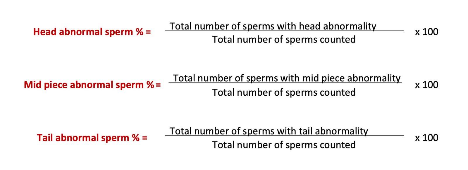
Interpretation of Result
Inference
- A maximum of 20% sperm abnormalities are allowed in bull semen (major 7.5% and minor 12.5%).
- Hereditary defects should not exceed 5%.
- Specific minor abnormalities should not exceed 10%.

