TABLE OF CONTENTS
Phimosis and Stenosis of the Preputial Orifice in Male Animals
Phimosis is the inability to protrude the penis beyond the sheath or prepuce. It may be congenital in young dogs, cats, and horses.
Stenosis of the preputial orifice is usually acquired due to injuries, wounds, and infections.
Etiology
In cattle with pendulous sheaths, the preputial orifice may be stepped on, causing severe contusion and swelling.
Chronic prolapse of the prepuce is a very common cause of posthitis and phimosis. It is especially common in Bos indicus cattle.
Pendulous sheath, large preputial orifice, and relaxed preputial membranes are inherited traits.
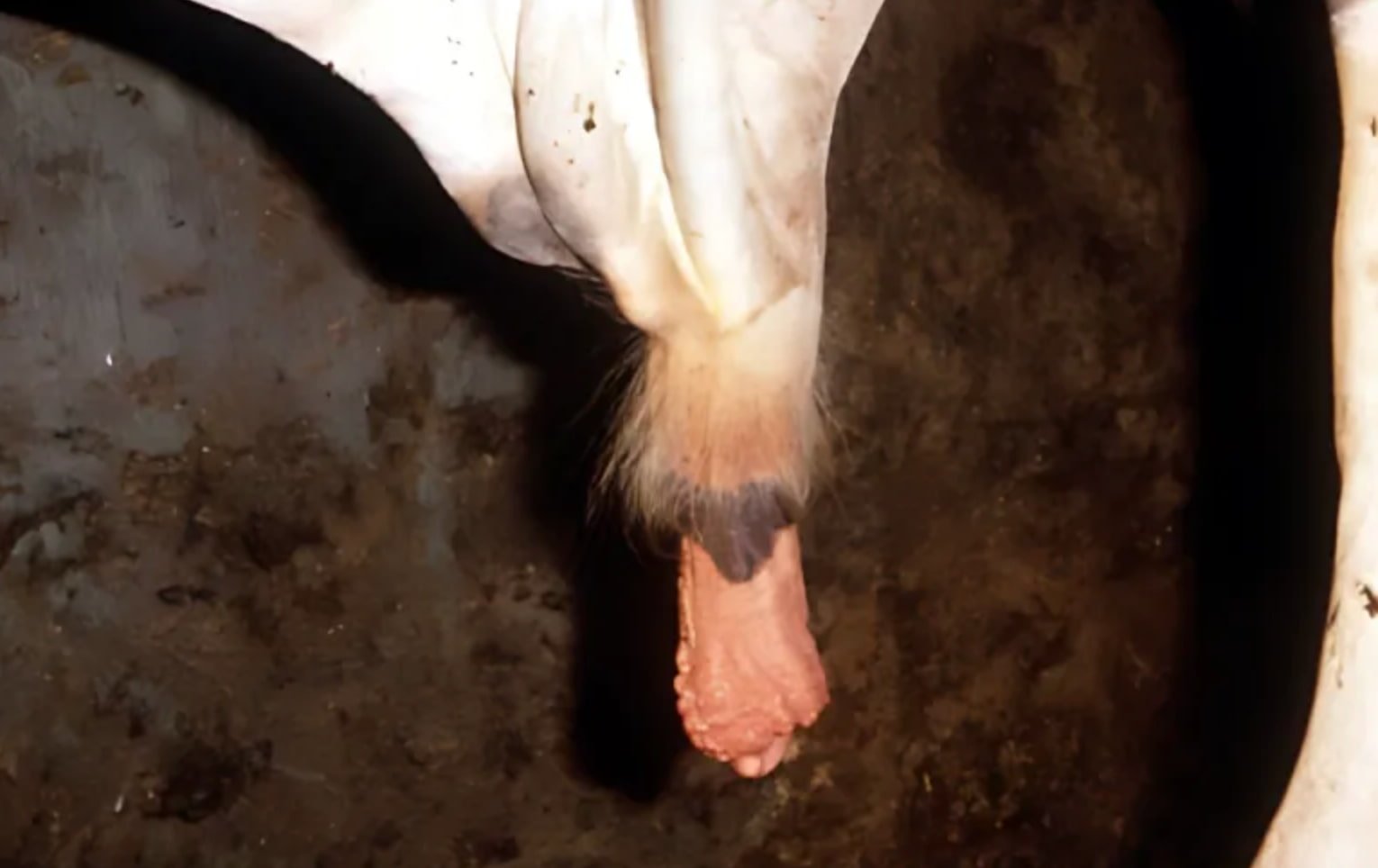
In affected breeds, prolapse of the prepuce may occur, and it becomes dry, traumatized, swollen, and fibrotic.
Predisposing factors are:
- Balanoposthitis
- Congenital adhesions
- Papillomata
- Prolapse of the prepuce
Management
In mild cases, the affected bull may be confined. The prolapsed organ should be carefully washed, cleaned, and dried.
Congenital stenosis in dogs may be corrected by a dorsal incision of the external preputial orifice.
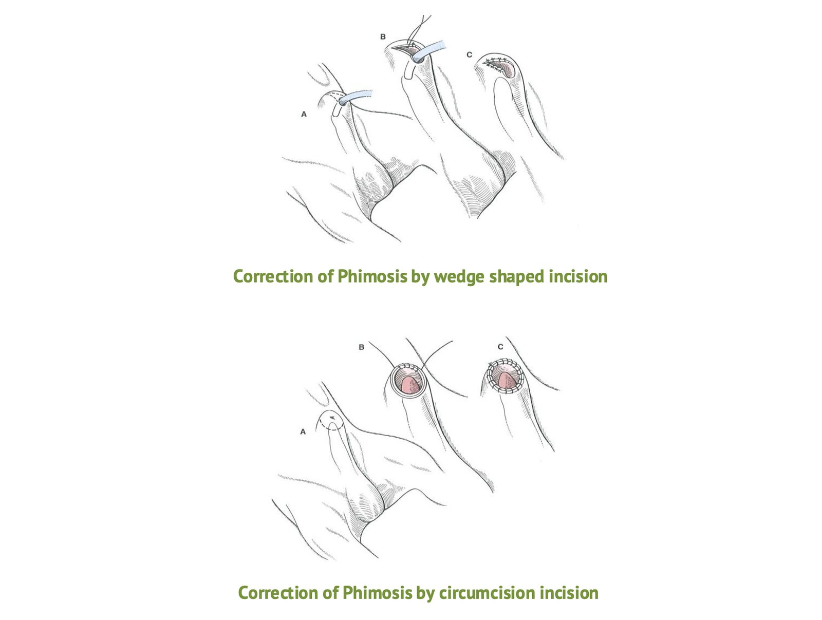
In bulls, dogs, or rams, the usual procedure to correct simple stenosis of the preputial orifice caused by cicatricial tissue is to remove a triangular portion of skin from the ventral part of the sheath or prepuce.
The base of the triangle is at the preputial orifice.
After the skin is removed, an incision is made through the midline of the prepuce to the apex of the triangle.
After careful hemostasis, the preputial membrane is sutured to the skin using interrupted catgut sutures.
Oily antibiotics or bland antiseptic preparations are applied, and the prolapsed organ is replaced and held in position by a purse-string suture through the preputial orifice.
In more severe cases where replacement is not possible, circumcision or even amputation of the prepuce is necessary.
Prevention
It is undesirable to operate on an affected bull since prolapse of the prepuce is genetically predisposed.
Males should be selected for lighter, less pendulous sheaths, smaller preputial orifices, and stronger retractor penis muscles.
Surgical Procedure
The surgery necessary for the correction of this condition consists of the removal of the diseased portion of the prepuce.
Strict asepsis and hemostasis are necessary for successful surgery.
A triangular portion of skin is removed from the ventral side of the sheath, with the base of the triangle including the margins of the original orifice.
The apex of the triangle is posterior to the orifice and lies on the midline of the bull.
The incision is carried through the preputial membrane, and the prepuce is joined to the margin of the skin by interrupted nylon sutures.
Care should be exercised to anchor the prepuce to the skin in several places before the diseased portion is removed to prevent the bull from drawing the penis and free edge of the prepuce deep into the sheath.
Post-operative strictures of the orifice may redevelop, despite generous V-shaped incisions and careful techniques.

