TABLE OF CONTENTS
Marek’s Disease
Marek’s Disease also known as Range paralysis, Skin leukosis, Neural lymphomatosis and Gray eye disease in animals.
Marek’s disease is a lymphoproliferative disorder of chicken is characterized by oncogenic transformation of T lymphocyte that infiltrate into lymphoid and other visceral organs.
Etiology
- Marek’s Disease is caused by Alphaherpesvirus, belongs to family Herpes viridae, which contain 3 serotypes
- Serotype 1 – oncogenic, serotype 2 &3 is non oncogenic in nature.
- The serotype 3 is called as turkey herpes virus (HVT).
- Serotype 1 contain variable pathotypes namely mild (m), virulent (v), very virulent (vv) and very virulent plus (vv+).
- The virus is stable in an environment and inactivated by a variety of common chemical disinfectants within ten minute treatment period.
- All the herpes viruses are sensitive to ether. The infectivity of dried MDV infected feather is destroyed by chlorine, organic iodine, quaternary ammonium compound, cresylic acid, synthetic phenol and sodium hydroxide.
Epidemiology
- Distribution: Worldwide.
- Chicken, turkey and quail are commonly affected.
- The disease most commonly occur between 12- 30 weeks of age.
- White leghorn breeds are highly susceptible than other breeds.
- Transportation, vaccination, handling and beak trimming are stress factors that increase susceptibility to diseases.
- The concurrent infection with Infectious bursal disease (IBD), Chicken infectious anemia (CIA) and presence of high level of aflatoxin in feed increases the susceptibility to disease.
Source of infection
- Feather follicle, dander, litter, the virus is infective for several months.
- Infected birds become carrier for life and remain source of infection by excretion of virus through feather follicle.
Transmission
- Inhalation of virus from feather follicle, dander and dust in poultry house environment.
- Darkling beetle can transmit the virus mechanically.
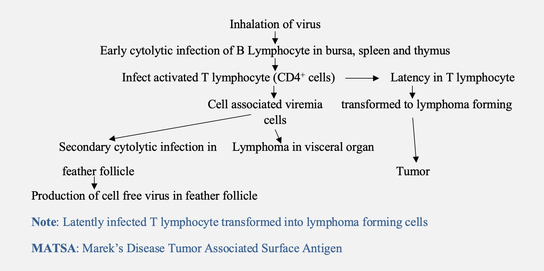
Pathogenesis
Clinical manifestation
- It occur as classical form and acute form.
Classical form
- Partial or complete paralysis of leg and wings results in stretching of one leg forward and another backward.
- Torticollis occur when nerve controlling muscle is affected.
- Involvement of vagus nerve results in paralysis and dilatation of crop. These bird shows symptom of gasping and respiratory distress.
Acute form
- Usually lymphoma formation in visceral organ.
- Generalized signs are depression, weight loss, anorexia and diarrhoea.
- Mortality is 10-30 % and in major outbreaks can go upto 70%.
Acute cytolytic disease
- Infection caused by very virulent plus strain.
- Severe atrophy of lympnoid organs.
- Acute cytolytic form also called as early mortality syndrome.
- Mortality is high usually occur between 10-14 days of age.
Transient paralysis
- Uncommon manifestation of MDV infection occur between 5-18 weeks of age.
- Sudden development of varying degree of ataxia, paresis or paralysis of the legs, wings and neck.
Immunosuppression
- Loss of lymphocyte as a consequence of virus replication.
- Virus induced changes in regulation of immune responses.
- Tumour cell induced immunosuppression.
Clinical manifestation of Marek’s disease in different forms:
Ocular form
- Blindness in birds due to mononuclear cell infiltration in the iris causing “grey eye” or “pearl eye”.
Skin or cutaneous form
- Distinct white nodules on the skin and in extreme cases looks like brownish nodules.
Muscular form
- Superficial and deep muscles like pectoral muscles affected.
- Whitish grey and there are tiny white streaks to nodular tumours in the muscles.
Necropsy Finding
- Enlargement of vagus, brachial or sciatic nerve, loss of striations and glistening nerve.
- Nodular lymphoid tumour in liver, spleen, gonads, heart, lung, kidney and proventriculus and feather follicle of skin.
- Acute cytolytic form- extensive atrophic changes in the thymus and bursa of fabricious may result in complete disappearance of the thymus and bursa.
Sample collection
- Feather follicle, buffy coat and visceral organ (tumor growth).
Diagnosis
- Based on clinical symptoms and necropsy finding.
- Histopathological examination- pleomorphic infiltration of lymphocytes and presence of MD cell in liver and spleen.
- The infection is usually detected by inoculating live buffy coat cells on to monolayer cultures of chicken kidney cells or duck embryo fibroblasts, in which characteristic viral plaques develop within a few days.
- Detection of antigen by AGID and PCR.
- Detection of antibody by AGID.
Differential diagnosis
- Lymphoid leukosis
Treatment
- There is no specific treatment.
Prevention and control
- Vaccination of day old chicks with either monovalent (HVT) or bivalent (HVT+SB-1) live vaccine with correct dose.
- CVI988/Rispen strain used to protect against very virulent plus (vv+) strain of serotype 1
- In ovo- vaccination of chicks at 18 days of embryonation.
- Proper handling, thawing and reconstitution to ensure adequate dose to be administered.
- Recombinant vaccine is available for fowl pox and Marek’s disease virus.
- Recently, marek’s disease virus lacking meq oncogene l confer protection against challenge with vv+ strain of MDV.
- Adequate cleaning and disinfection of poultry house before introduction of chicks.
- Rearing of birds in all –in all-out system.
- Selection of genetically resistance strain.
- Avoid feeding of aflatoxin level in feed above permissible level (>0.02 ppm).

