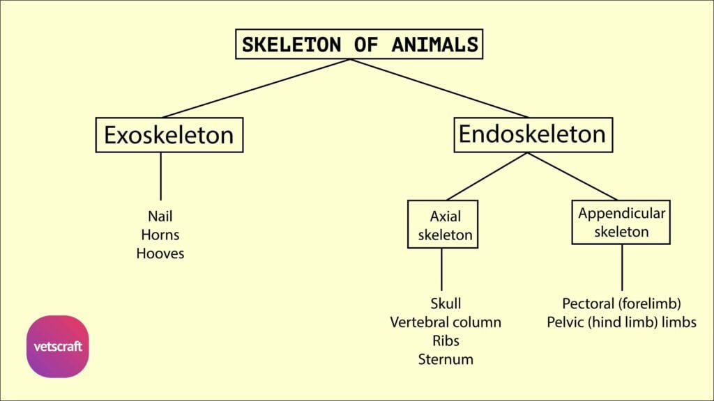
TABLE OF CONTENTS
Mammary gland or udder of Animals
The mammary glands are modified cutaneous gland associated functionally with the genital organs. They are popularly known as “Udder”.
Mammary gland of Cow
- Mammary gland or udder of cow is compound saccular glands, four in number and two on the either side of the median line
- They extend from caudal part of the abdomen to the floor of the pelvis and thus lie between the thighs
- Each gland is flattened from above downwards and presents a base and an apex and two lateral faces
- The lateral face is convex and the medial surface is flattened. The base is slightly concave and slopes downwards and forwards in adaptation to the abdominal wall
- The appearance of the udder varies greatly, depending on maturity and functional status of the individual and also based on the breed character
- The base of each gland is attached to the abdominal wall by means of a well-developed suspensory apparatus, which is attached to the ventral part of pelvic symphysis by means of the subpelvic tendon. This sub-pelvic tendon attaches the prepubic tendon to the ventral part of the pelvic symphysis
- The suspensory apparatus (ligament) consists of four sheets of tissue.
- Of which two are median and two are lateral in position. The median laminae are well developed and median in position and are chiefly made up of yellow elastic tissue
- The two glands of either side are separated by the double septum, which attaches to the medial flat surface of the gland
- The lateral laminae are made up of dense connective tissue
- It arises from the subpelvic tendon caudal to the udder
- It descends downwards, while reaching the abdominal floor they diverge into median and lateral laminae
- The lateral laminae then extend downward over the udder and divide into superficial and deep layers
- The superficial layer attaches to the skin where it reflects off the udder to the medial face of the thigh
- The deep layer is thicker and attached to the convex lateral surface of the udder by numerous lamellae, which pass into the gland
- The caudal part of the base has the supramammary lymph gland and a considerable amount of fat in relation to it
- It is customary to divide the udder into four quarters. There is neither septum nor visible division between the two quarters of the same side
- Each quarter is independent of the other as each is a compound saccular gland
- A teat continues the apex of each quarter, which is about 7-8 Cm long
- A prominent inter mammary groove marks the division of the udder into right and left halves. A single teat continues the apex of each quarter
- Each teat has a single lactiferous duct (Ductus lactiferous), which widens superiorly into a roomy lactiferous sinus (Sinus lactiferous), popularly known as milk cistern
Mammary gland of Ewe and doe
Mammary gland of Ewe and doe are two in number and relatively large. They are approximately globular but flattened on the septal side.
Mammary gland of Mare
There are only two Mammary glands present in mare, which are not divided into quarters smaller than those of the cow, each gland has a single teat and the apex presents two openings close together. Two lactiferous ducts lead into the sinus. Each teat is 2.5 Cm – 5 Cm long.
Mammary gland of Sow
Mammary gland are usually ten or twelve in number in sow and are arranged in two rows as in the bitch. Each teat has commonly two ducts.
Mammary gland of Bitch
The Mammary glands are 10 in number in bitch arranged in two series of five each as pectoral, abdominal and inguinal. The apex of each teat has 6-10 openings on it.
Mammary gland of Rabbit
Four or five pairs of nipples present in rabbit on the ventral surface of the abdomen and thorax. The lactiferous sinus is not prominent.