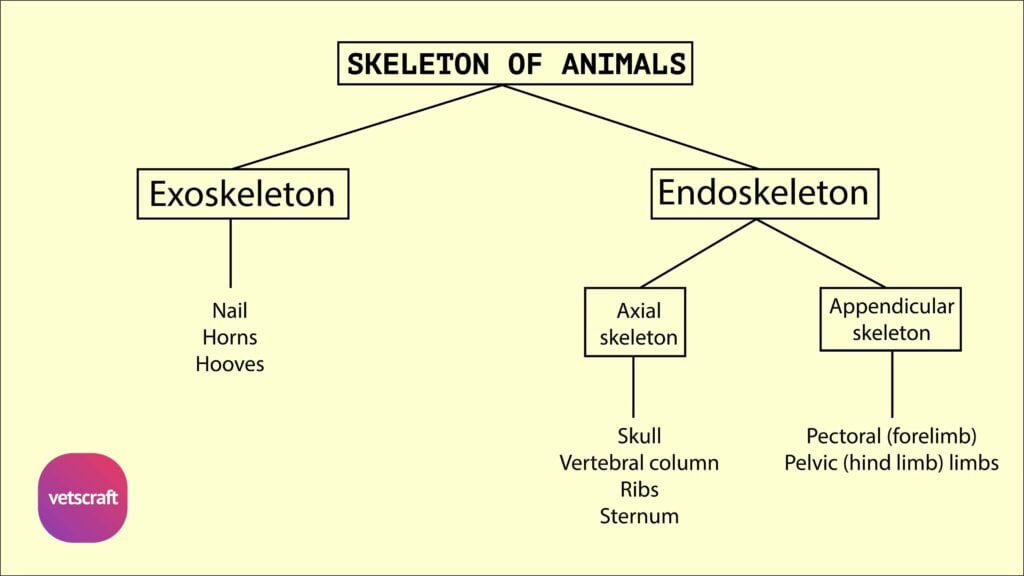
TABLE OF CONTENTS
Interior of Ruminant Stomach of Ox
Interior of Ruminant Stomach of Ox different compartments of the ruminant stomach differ in order to accomplish its function.

Rumen
- The cavity of the rumen is divided into two sacs by the pillars of the rumen, which are muscular folds and correspond to the grooves on the exterior
- They project like shelves into the cavity of the organ. The cranial pillar has a thick concave free edge
- Between these free edges, the two sacs of rumen communicate with each other
- The right and left pillars connect the cranial and caudal pillars and are less prominent
- The rumino-reticular fold corresponds to the rumino-reticular groove. Its free edge is concave and forms the ventral and lateral margins of the large oval rumino-reticular aperture
- The cardia is about 10 to 12 cm ventral to the vertebral end of the 8th or 9th rib. The opening is slit-like
- The mucous membrane is brown in colour except on the pillars where it is pale
- It is thickly studded with papillae which are however not present on the pillars.
OESOPHAGEAL GROOVE

The reticular or oesophageal groove is formed by two muscular ridges or lips extending from the cardia to the reticulo -omasal opening.
- It is about 18 to 20 cm length. Its direction is chiefly dorso-ventral but usually it inclines somewhat forward and medially in its ventral part
- The groove is twisted in spiral fashion so that its thickened edges project at first backward then to the left and finally forwards
- The twist mainly concerns the left lip
- The two lips meet dorsal to the cardiac opening and pass ventrally along the right wall of the reticulum, the right lip twisting around the left lip
Reticulum
- The interior of the reticulum is raised into folds of about 1/2 inch high enclosing 4 to 6 sided spaces or cells.
- Smaller folds subdivide these cells and bottoms are studded with pointed horny papillae
- The reticulo-omasal orifice is situated at the lesser curvature of the reticulum five or six inches above the bottom of the later and is rounded
Omasum
- The cavity of omasum is occupied by about hundred longitudinal muscular folds – the lamina omasi, which spring from the greater curvature
- The largest of these are about half a dozen in number, have a superior convex attached edge and a thick concave free edge
- A groove sulcus omasi extends from the reticulo-omasal opening to the omaso-abomasal opening and is about 4 inches long
- It is free from laminae. The omaso-abomasal orifice is oval and is about 4 inches long
Abomasum
- The abomasum is an elongated sac, which lies on the abdominal floor from the xiphoid cartilage backwards
- The cranial blind end is at the xiphoid region in relation with the reticulum
- The body extends back between the ventral sac of rumen and the omasum and turns to the right behind the omasum
- It is constricted about the middle forming an cranial larger part and a caudal pear shaped smaller part
- The pyloric part inclines dorsally and joins the duodenum at the ventral part of the 10th rib
- The parietal surface is in contact with the abdominal floor. The visceral surface is related to the rumen and omasum
- The greater curvature gives attachment to the superficial part of the greater omentum
- The lesser curvature is related to the greater curvature of the omasum.