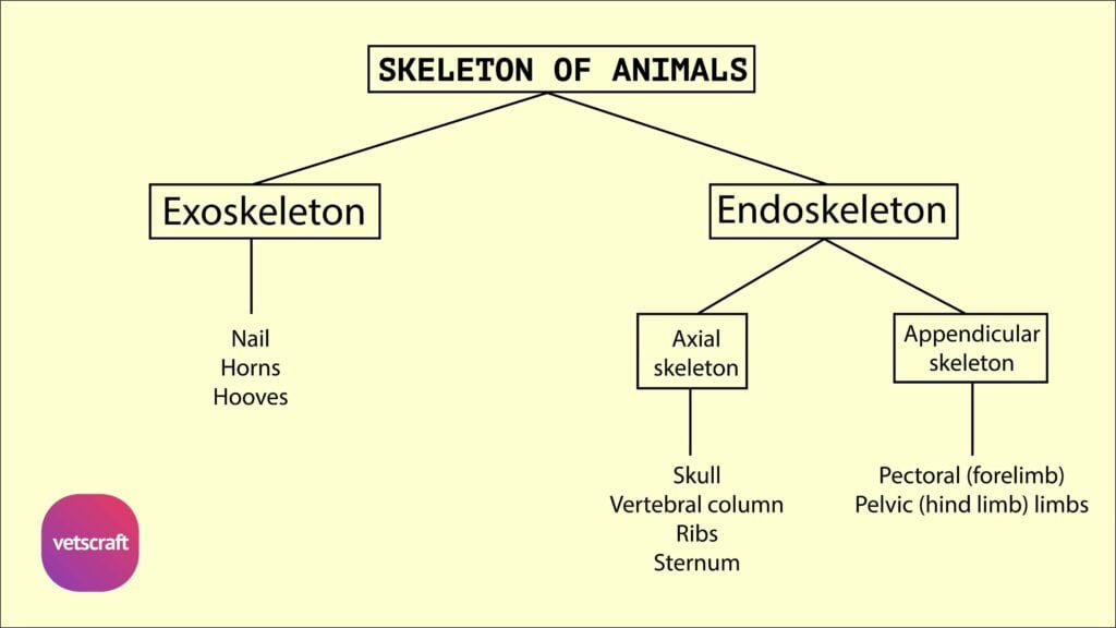
TABLE OF CONTENTS
Comparative Anatomy of the Humerus Bone in Animals
The humerus is a long bone located in the forelimb of animals. It extends obliquely downward and backward between the shoulder joint (proximally) and the elbow joint (distally).
The humerus serves as a crucial structure for limb movement, muscle attachment, and joint articulation.
Humerus Bone of Ox
The humerus bone of the ox is a long bone positioned obliquely downward and backward between the shoulder joint above and the elbow joint below. It has a shaft and two extremities.
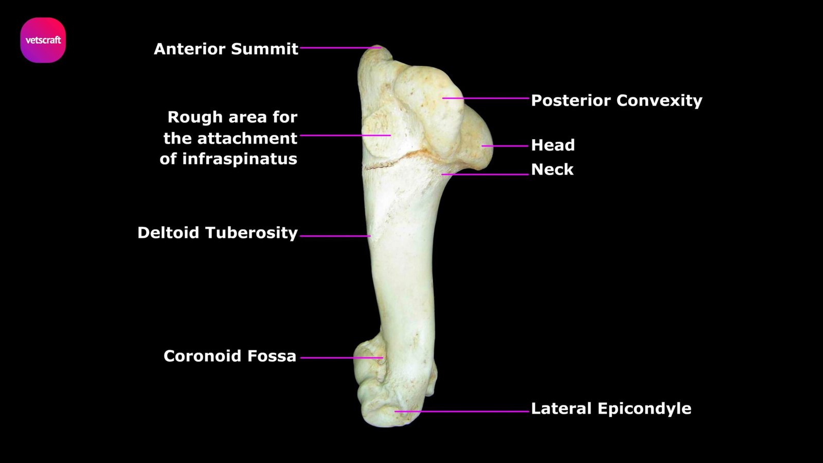
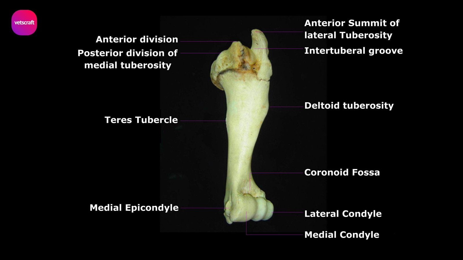
Shaft
The shaft of the humerus of ox has a twisted appearance and has four surfaces. The anterior face is triangular, wide and smooth above, narrow and rough below.
The posterior face blends with the medial and lateral faces. It presents the nutrient foramen about the middle.
The medial face is nearly straight in its length. Just above its middle, it presents the teres tubercle for latissimus dorsi and teres major.
The lateral face is spirally curved and forms the musculo-spiral groove, which contains the brachialis muscle. The groove is continuous with the posterior face above and winds around towards the front. The lateral surface is separated from the anterior by a distinct border, the crest of the humerus, which bears above its middle the deltoid tuberosity. The crest is for the insertion of brachiocephalicus and superficial pectoral, and the deltoid tuberosity for the deltoideus muscle. A curved line extends upward from the deltoid tuberosity and is for the lateral head of triceps. At the upper part of the curved line is a nodule for the teres minor.
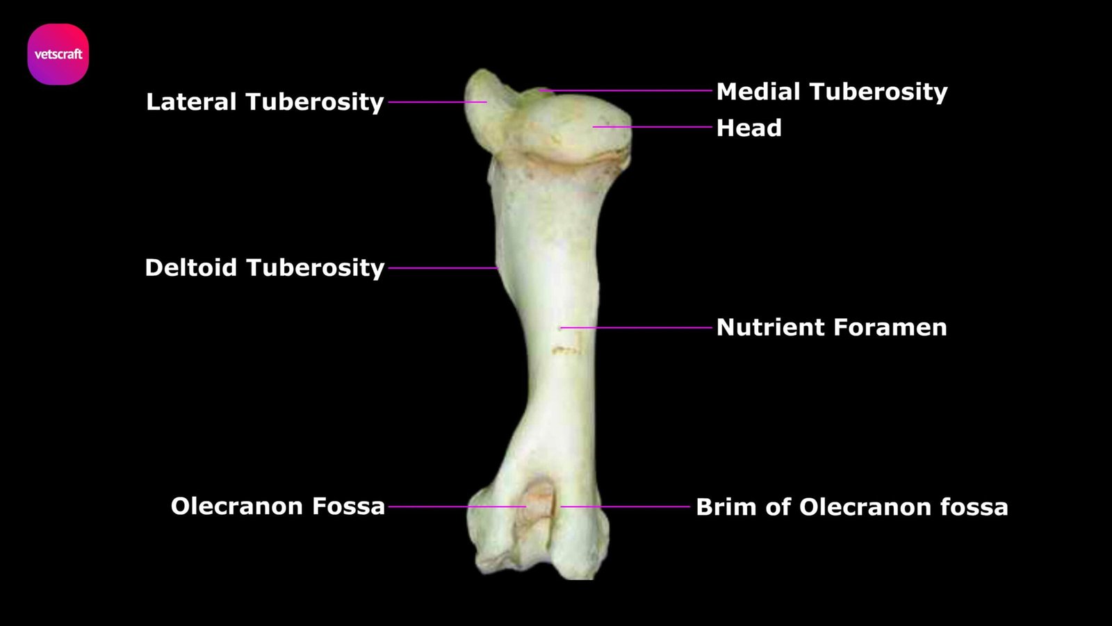
Proximal Extremity
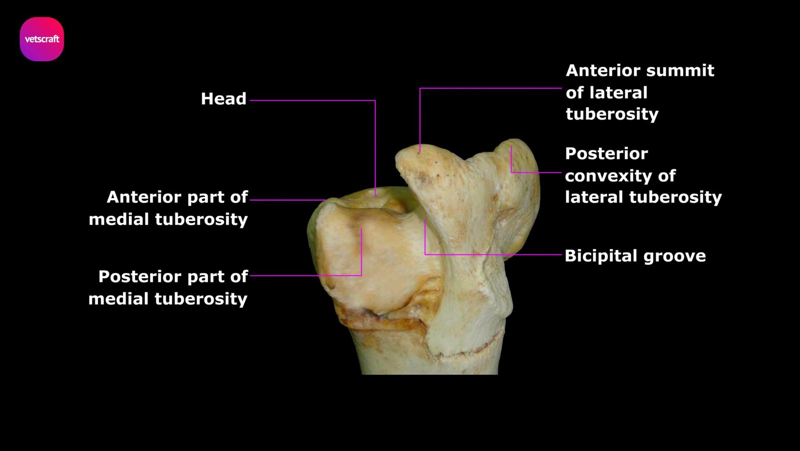
The proximal extremity consists of a head, neck, two tuberosities, and the intertuberal or bicipital groove. The head presents a circular articular surface, which articulates with the glenoid cavity of the scapula to form the shoulder joint.
The lateral tuberosity consists of two parts—an anterior summit arching medially and a posterior convexity. The former overhangs the bicipital groove and gives attachment to the lateral tendon of the infraspinatus. The convexity gives attachment medially to the medial tendon of the infraspinatus.
The medial tuberosity is smaller and consists of anterior and posterior parts. The anterior part forms the medial boundary of the intertuberal groove and gives attachment to the medial tendon of the supraspinatus and to the deep pectoral. The posterior part is for the subscapularis.
The intertuberal groove is bounded by the anterior divisions of the two tuberosities and, in life, is covered by a fibrocartilage for the play of the tendon of origin of the biceps brachii.
Distal Extremity
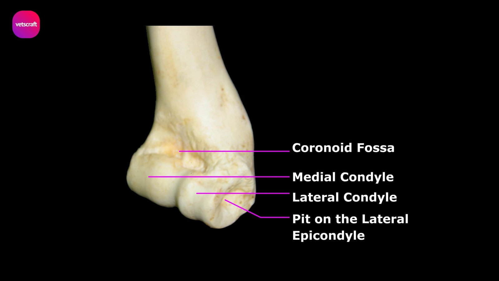
The distal extremity has an oblique surface, which is divided by a ridge into two condyles, the medial being the larger. The medial condyle is crossed by a groove, which extends into the olecranon fossa and articulates with the semilunar notch of the ulna.
This extremity presents, anteriorly above the articular area, the coronoid fossa, which receives the coronoid process of the radius in extreme flexion of the elbow joint.
Behind and above the condyles are two thick ridges—the epicondyles. The medial epicondyle is more prominent and furnishes origin to flexor muscles of the carpus and digits and bears a tubercle for the attachment of the medial ligament of the elbow joint. The margins of the olecranon fossa are for the origin of the anconeus.
The lateral epicondyle bears laterally the condyloid crest in the form of a raised ridge, which gives origin to the extensor carpi radialis. Between these two epicondyles is the deep olecranon fossa, which receives the anconeus process of the ulna during extreme extension. The distal extremity also presents a rough depression laterally and a tubercle medially for the collateral ligaments of the elbow joint.
Humerus of Sheep and Goat
The humerus of sheep and goat is relatively longer and more slender than that of the ox. The deltoid tuberosity is closer to the proximal end and not as prominent.
The lateral tuberosity is smaller and does not overhang the bicipital groove. The olecranon and coronoid fossae are shallower.
Humerus Bone of Horse
The deltoid tuberosity is more prominent in the humerus of the horse. The bicipital groove is divided by an intermediate ridge.
The summit of the lateral tuberosity does not arch inward. The coronoid and olecranon fossae are shallower.
Humerus of Pig
The humerus bone of the pig has the appearance of an italic letter ‘f’ minus the crossbar. The shaft is laterally compressed and flat on the medial side.
The musculo-spiral groove is shallow, and the deltoid tuberosity is small. There is a larger, rounded eminence midway between it and the lateral tuberosity.
Other features of the pig’s humerus:
- The teres tubercle is absent.
- The condyles are of equal size.
- The coronoid fossa is prominent.
- The olecranon fossa is narrow and deep.
- Occasionally, the supratrochlear foramen is present.
Humerus Bone of Dog
The musculospiral groove is shallow, and the deltoid tuberosity is in the form of a low ridge in the case of the dog. The lateral tuberosity is undivided. The coronoid and olecranon fossae communicate with each other through the supratrochlear foramen, through which no soft structure passes.
Humerus of Rabbit
The head of the humerus in the rabbit is globose, and at the distal extremity, the articular surface is divided into two parts by a ridge.
Humerus of Fowl
The humerus bone of the fowl is directed parallel to the thoracic vertebrae when the wing is at rest. The proximal extremity presents, on its medial aspect, an opening leading into the air cavity within the shaft.