TABLE OF CONTENTS
Fetotomy or Embryotomy in Veterinary Obstetrics
Fetotomy, also known as embryotomy, is a crucial procedure in veterinary obstetrics, especially in managing dystocia cases. This guide explores its types, advantages, disadvantages, and procedure in large animals.
Fetotomy or embryotomy is defined as an operation performed on the fetus to reduce its size by either dividing it or removing certain parts.
Fetotomy is used most commonly in cattle, occasionally in horses, rarely in sheep and goats, and almost never in pigs and small animals.
Types of Fetotomy
Partial Fetotomy or Incomplete Fetotomy
When a small part of the fetus is removed, the operation is known as incomplete fetotomy.
Total Fetotomy or Complete Fetotomy
When the whole fetus is divided into small pieces, the operation is known as complete fetotomy. In fetotomy, the life of the fetus is sacrificed if it is not already dead.
Advantages of Fetotomy
- It reduces the size of the fetus.
- It avoids caesarean operation.
- It requires little assistance.
- It will prevent possible trauma or injury to the dam through the use of excessive traction.
Disadvantages of Fetotomy
- It may be dangerous, causing injuries or lacerations to the uterus or birth canal by instruments or sharp edges of bone.
- It may take a long time, exhausting both the dam and the operator.
- It is conducive to trauma and pressure necrosis of the birth canal.
- It offers certain dangers to the operator by wounds from instruments, and if the fetus is emphysematous there is a possibility of infection of the operator’s arm.
Fetotomy Operations
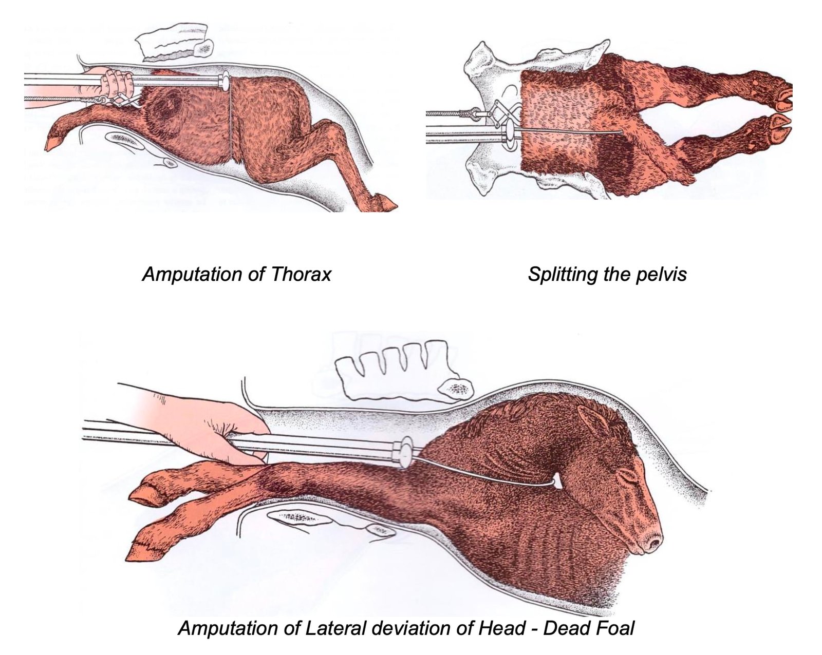
Decapitation or Amputation of the Protruding Head
Decapitation or amputation of the protruding head is indicated when one or both forelimbs are retained and the head has passed through the vulva and become swollen, edematous, or emphysematous, making it impossible or extremely difficult to repel.
In such cases, the fetus is usually dead, and amputation of its head allows the fetus to be repelled, enabling correction of the abnormal posture of the forelimbs through mutation without interference from the head.
Cephalotomy or Craniotomy
Cephalotomy or craniotomy is indicated when fetal hydrocephalus or other cranial deformities are the cause of dystocia.
Amputation of the Head and Neck
Amputation of the head and neck is indicated when the head and neck are retained alongside, beneath, or above the body of the fetus.
It may also be indicated when, due to a juvenile or hypoplastic pelvis, or a large fetal head, there is insufficient space in the birth canal for the simultaneous presence of the head, neck, and both forelimbs.
Amputation of the head and neck may be performed using a wire saw or fetotome.
Amputation of the Forelimb
There are two types of operations:
- Subcutanesous fetotomy: Removal of decorticated limb (skinless limb) to reduce the size of the fetus.
- Percutaneous fetotomy: removal or division of certain parts of fetus along with intact skin to reduce the size of the fetus.
1. Subcutaneous Fetotomy
Subcutaneous fetotomy is relatively simple and rapid, especially when the fetus is emphysematous. It consists of applying steady traction to the limb using a snare placed around the fetlock, then making an incision at the top of the scapula with a knife, cutting through the skin and into the muscle along the entire length of the leg down to the pastern.
The operator uses the thumb and fingers to separate the skin from the underlying limb tissue, starting at the fetlock joint.
The skin is then detached from the leg at the fetlock by cutting through the joint, so that the pastern bones and hoof remain attached to the skin. Strong traction is applied to the limb using the snare around the metacarpal bone.
The pectoral muscles, nerves, and arteries beneath the scapula are broken manually with the fingers or severed using a knife. Traction usually separates the muscles at the dorsal end of the scapula; if not, these muscles must be incised.
2. Percutaneous Fetotomy
Amputation of the forelimb is initiated by applying constant traction to the leg using a snare or obstetrical chain.
A crescent-shaped incision is made through the skin of the fetus and its trapezius and rhomboideus muscles, dorsal to the scapula, using a concealed knife. This separates the dorsal end of the scapula from the body. A loop of the fetotome is placed in the pectoral region. The wire loop is then moved upward and positioned in the incision under the cartilage of the scapula, and the limb is removed.
The removal of a forelimb extended beneath the body is easily performed by passing the fetotome wire behind the elbow and beneath the arm. By placing the head of the fetotome on the withers of the fetus, the entire limb can be removed rapidly.
Evisceration in Anterior Presentation
Evisceration in anterior presentation is indicated to reduce the size of a large or emphysematous fetus or to release the ascitic fluid in fetal ascites.
When the fetus is within the birth canal or uterus, evisceration in anterior presentation may be performed by amputation of the forelimb with a fetotome, which exposes the ribs.
The intercostal muscles between the ribs are separated by the finger or knife from the spine to the sternum. Then the heart and lungs are removed by manual traction after grasping the heart at its base and grasping the fingers firmly around the large vessels. By grasping the lungs in the region of the bifurcation of the trachea, first one and then the other or both lungs can be removed.
After the thoracic viscera have been removed, the fingers are thrust through the diaphragm into the abdomen. The stomach and intestines are easily removed by wrapping them around the hand.
The liver, the largest organ to be removed, is pulled loose from the diaphragm and withdrawn, usually in several pieces. The kidneys are small and therefore need not be removed.
Detruncation in Anterior Presentation
Detruncation in anterior presentation is indicated in rare instances when the fetus is in a dog-sitting posture with the hind limbs extended alongside or beneath the body and wedged into or against the maternal pelvis.
A loop of fetotome wire is passed around the body of the fetus, and the head of the fetotome is inserted into the birth canal over the lumbar region of the fetus. After the wire is manipulated on the back of the last rib, detruncation is rapidly accomplished.
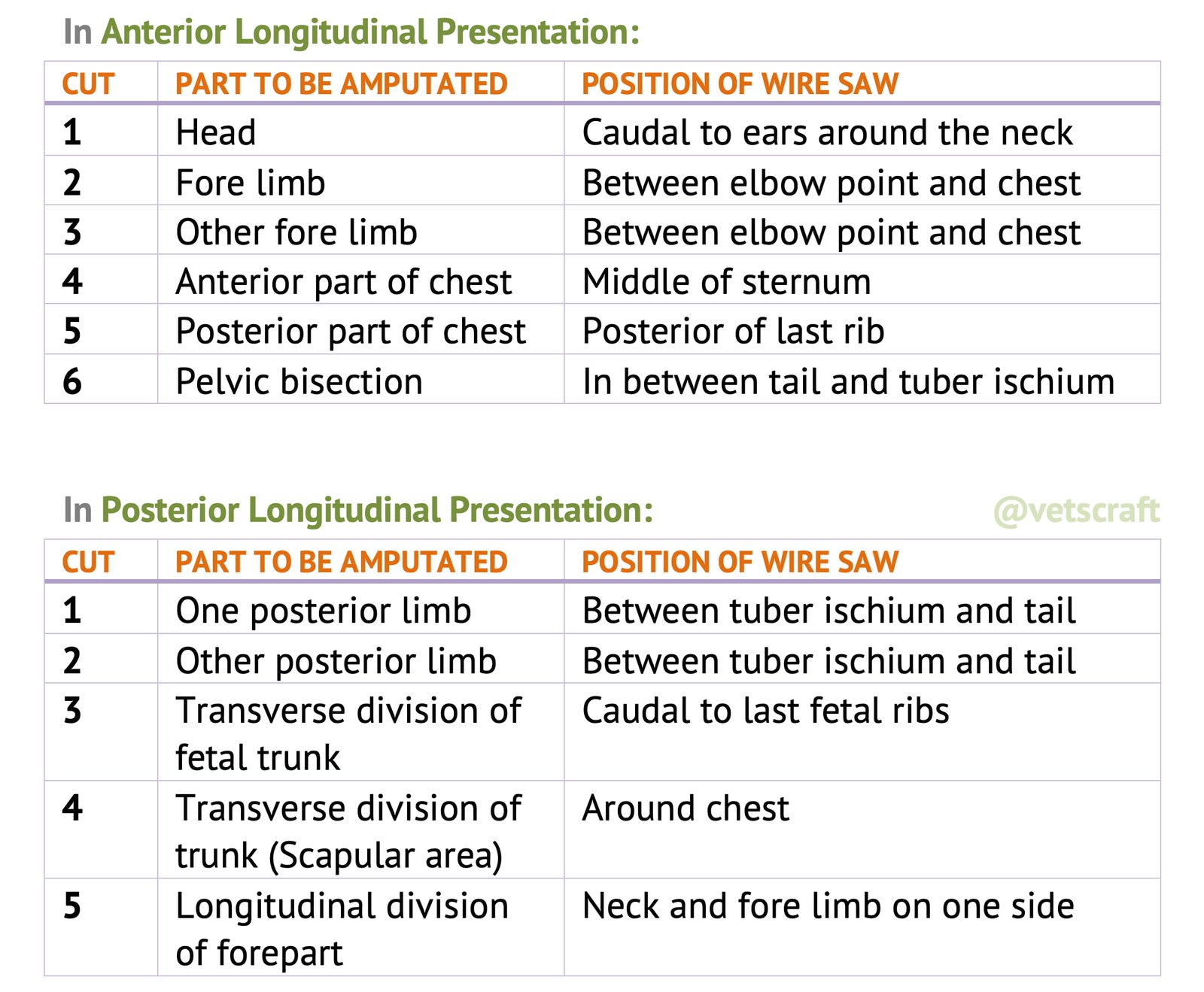
Bisection of the Pelvis in Anterior Presentation
Bisection of the pelvis in anterior presentation is indicated when the fetal pelvis becomes wedged in the maternal pelvis. This may be performed using a fetotome or wire saw.
Bisection of the Pelvis in Breech Presentation
Bisection of the pelvis in breech presentation is indicated in posterior presentation in uniparous animals when the hind limbs of the fetus are extended or retained beneath the body. This condition often occurs after prolonged dystocia, leading to uterine contraction and fetal emphysema.
In such cases, the uterus and birth passages become dry and swollen, making mutation difficult or impossible. The operation can be performed using a wire saw or a fetotome to facilitate fetal removal.
Evisceration in Posterior Presentation
After the removal of one or both of the posterior limbs the opening into the abdominal cavity may be enlarged and the viscera removed as described under evisceration in anterior presentation but in a reverse manner, removing first the abdominal and then the thoracic viscera.

FAQs
Fetotomy, also known as embryotomy, is the dismemberment of a dead fetus inside the uterus to facilitate its removal when normal delivery is not possible.
Fetotomy is indicated in cases of dystocia due to a dead fetus, especially when the fetus cannot be delivered by traction and cesarean section is not preferred.
The main types are complete fetotomy, partial fetotomy, and subcutaneous decapitation, depending on the region and stage of fetal decomposition.
Instruments include the fetotome (e.g., Reindl type), OB wire, handles, OB chains, hooks, and protective gloves and lubricants for safe manipulation.
Proper restraint, ample lubrication, careful instrument handling, and continuous monitoring of maternal trauma risk are critical during the procedure.

