TABLE OF CONTENTS
Exocrine Pancreatic Insufficiency (EPI) in Small Animals
In Exocrine Pancreatic Insufficiency (EPI) in Small Animals (dog, cats etc.), the exocrine functioning of the pancreas is affected, which is clinically manifested by passing voluminous, glistening faeces, persistent weight loss in spite of good appetite, PICA, coprophagia (eating own faeces), etc.
Etiology
Exocrine pancreatic insufficiency (EPI) is rarely caused by pancreatitis; fibrosis of the gland leads to EPI. Idiopathic atrophy of the pancreas and adenocarcinoma of the pancreas. Signs are evident when 80% of the gland is destroyed. Treatment is symptomatic, and the prognosis is guarded.
Pathogenesis
Gland is destroyed, leading to a deficiency of pancreatic enzymes, resulting in maldigestion and malabsorption, causing osmotic diarrhoea and thus exhibiting symptoms of malabsorption.
Prolonged diarrhoea leads to the loss of vital elements like fat and water, soluble vitamins, and micronutrients, which are undigested and give a glistening appearance to the faeces. There is a loss of bicarbonate ions, which leads to acidification of the bowel, leading to damage to the intestinal mucosa. In due course, it may lead to inflammatory bowel disease (IBD).
Symptoms
- Frequent passing of large voluminous faeces (glistening and foul smelling).
- Animal has got very good appetite but persistent weight loss is seen (polyphagia).
- Coprophagia, pica, dull, dry hair coat, polyuria.
Diagnosis
- Based on clinical signs (passage of voluminous faeces, weight loss in spite of a good appetite).
- Faecal examination is the best tool: stool samples are tested for undigested starch, fat, and the presence of trypsin.
- Sudan III staining: A faecal smear is stained with Sudan III or IV. Plenty of yellow fat globules can be seen.
- Detection of trypsin in faeces: x-ray film is coated with gelatin, trypsin, being a proteolytic enzyme, digests gelatin. A strip of exposed x-ray film is dipped in the faecal solution (1:10) and kept in an incubator for 1-2 hours, and then the x-ray film is washed under tap water. If trypsin is present in the faecal suspension, the gelatin coat is cleared from that x-ray strip, which confirms the presence of trypsin in the faecal suspension.
- Starch: The faecal smear is stained with 2% tincture iodine. Blue coloured starch granules indicate a positive test.
- Plasma turbidity test: A specified quantity of fat meal is fed to the dog (corn meal is fed at 30 g/kg body weight). Plasma is collected 2, 3, and 4 hours post-feeding and checked for lipemia. Absence of lipemia indicates EPI.
Treatment
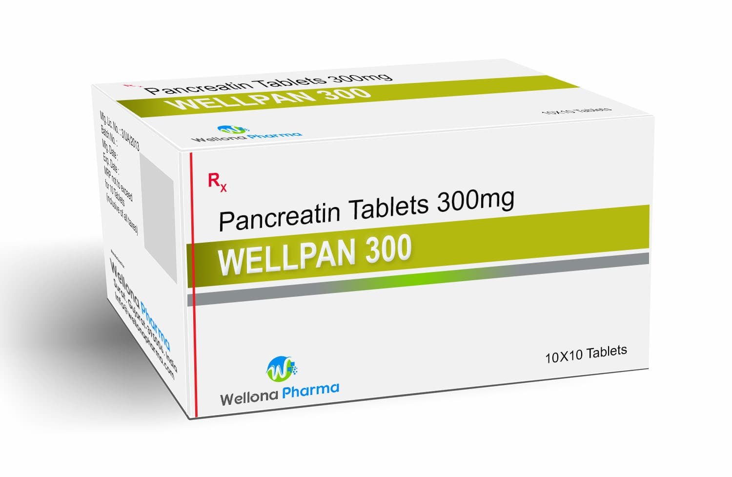
Exocrine Pancreatic Insufficiency (EPI) in Small Animals is treated by supplementation with digestive enzymes (Pancreatin tablets) and cobalamin.
Certain animals might not react to cobalamin treatment or enzyme supplements, and they probably also have small-intestinal sickness at the same time. Small-intestinal dysbiosis is frequently present in animals with EPI, and antimicrobial medication (such as tylosin) may be helpful. Certain animals with epilepsy also develop inflammatory bowel illness. It is also possible to try a proton pump inhibitor (Pantoprazole, Rabeprazole, etc.) for those who do not react to treatment.

