TABLE OF CONTENTS
Endocrine Regulation of Reproduction
The endocrine system plays an important role in regulating reproductive functions through the coordinated actions of hormones secreted by various glands, including the hypothalamus, pituitary, gonads, and placenta.
In contrast to the neural system, the endocrine system depends on hormones to cause the responses. Hormone in extremely small quantities can bring about dramatic physiologic responses.
Hormones are classified as having relatively short half lives. Hormonal half life is defined as the time required for one half of the quantity of the hormone to disappear from the blood or from the body.
Short half lives are important because once the hormone is secreted and released into the blood and cause a response, it is rapidly degraded so that further or unnecessary responses do not occur. However, when hormones such as progesterone are produced during pregnancy, the action brought about by the hormone continues as long as the hormone is present.
Compared to neural control, hormonal control is slower and has durations of minutes, hours or even days.
Mechanism Controlling Secretion
Positive and Negative Feedback are the major Controllers of Reproductive Hormones.
(1) Negative feedback
Inhibits GnRH neurons. For eg., High progesterone inhibits GnRH neurons which secrete only basal levels of GnRH. Such basal levels will allow for some follicular development but only to the extent where insufficient estrogen production is there. Hence, when progesterone levels are high, animal does not cycle. The tonic center in both and female is believed to respond mostly to negative feedback.
(2) Positive feedback
Activates the GnRH neurons in the hypothalamus. When estradiol reaches threshold level, the surge center will be positively stimulated and will release large quantities of LH that stimulate ovulation.
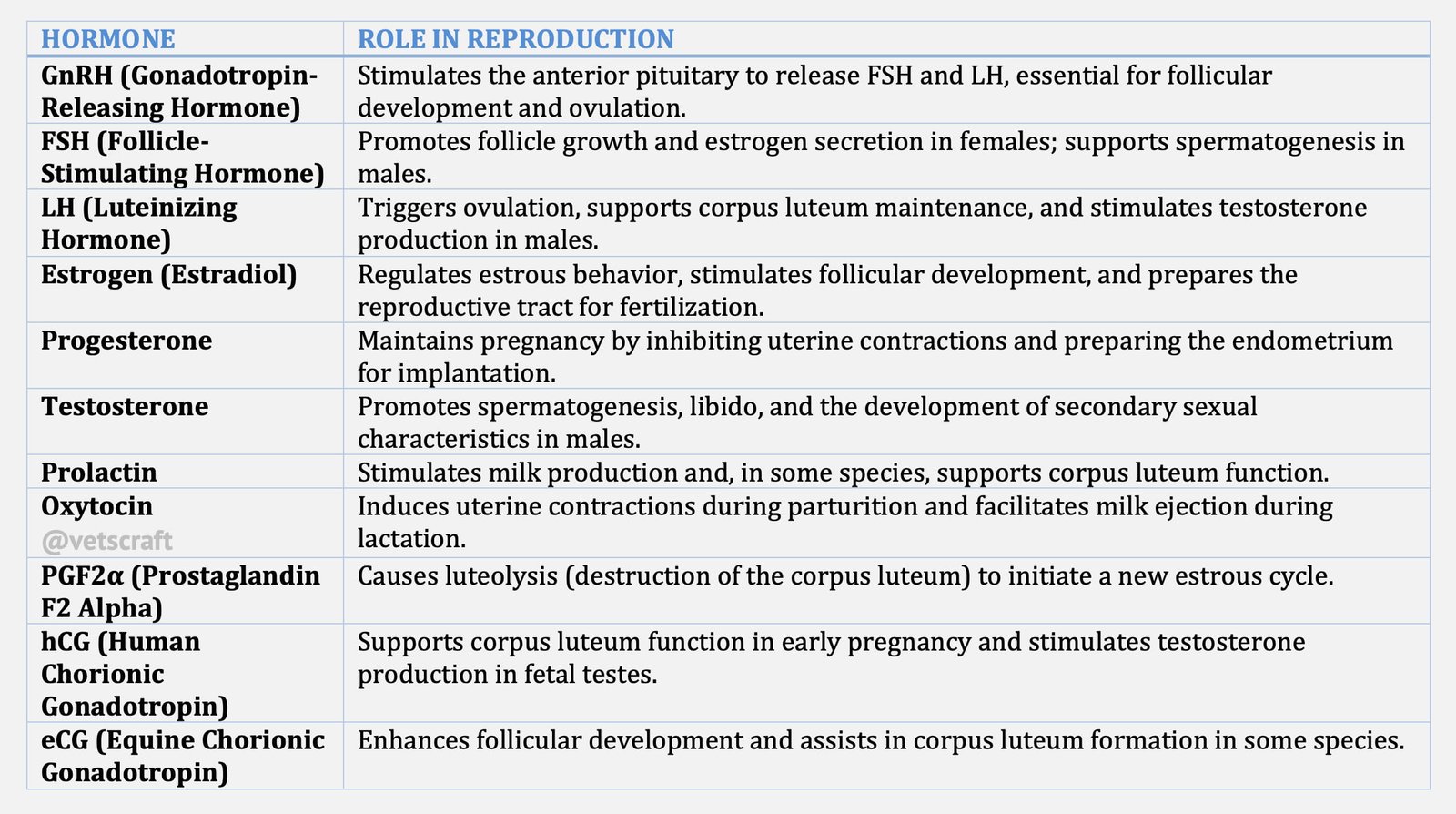
Hormones of Reproduction
Hormones of reproduction are classified based on:
- Chemical structure
- Glandular origin
- Mode of action
Chemical Structure
Based on their structure, the hormones are classified into:
- Proteins
- Steroids
- Fatty acids
Proteins or Polypeptide Hormones
Proteins or polypeptide hormones have molecular weight of 300 to 70,000 Daltons and are easily broken down by enzymes. They must be administered by injections.
They contain an α and β subunit. The α subunit for FSH, LH and TSH are identical but the β subunit is unique to each species. When two α or two β subunits combine the resulting molecule will have no activity. If α subunit of one hormone combines with the β subunit of another hormone, the activity of the molecule will be determined by the β sub unit only.
The amount of carbohydrate present on the surface of the protein determines the duration of half life of the hormone.
Glycoprotein hormones can be degraded easily by proteolytic enzymes in the digestive tract. There fore, they are not effective when given orally.
Steroids
Steroid hormones have the common cyclopentano perhydro phenantherene ring nucleus. They have a molecular weight of 300 to 400 Daltons.
Precursor is cholesterol. Natural steroids are not effective by oral administration.
Synthetic and plant steroids can be administered orally and by injection. Steroid hormones are sexual promoters and cause profound changes in both male and female reproductive tract.
Fatty acids
Fatty acids have a molecular weight of approximately 400 Daltons, and can be administered by injection.
Hormones & Glands in Reproduction
Reproductive hormones originate from:
- Hypothalamus
- Pituitary
- Gonads
- Uterus, and
- Placenta
Based on their origin, they are classified as:
- Hypothalamic hormones eg. GnRH.
- Pituitary Hormones eg. FSH/LH.
- Gonadal Hormones eg. Estrogen, Progesterone.
- Uterine hormones eg. PGF2 α.
- Placental hormones eg. hCG, eCG.
Mode of Action
- Neurohormones: Synthesized by neurons and released directly into the blood and cause response in a target tissue. eg. Oxytocin. A neurohormone can act on any number of tissues provided that the tissue has cellular receptors for the neurohormone.
- Releasing hormones: Synthesized by neurons in hypothalamus and cause release of hormones from pituitary. eg. GnRH.
- Gonadotrophins: Hormones released by the gonadotroph cells of the anterior pituitary and stimulate gonads. The suffix `tropin‘ means having an affinity for. eg. FSH and LH.
- Sexual promoters (Steroids): Produced by the gonads of both male and female to stimulate the reproductive tract, to regulate function of hypothalamus and anterior pituitary and to regulate reproductive behaviour.
- Pregnancy maintenance hormones: are responsible for maintenance of pregnancy. (eg. Progesterone) and in some cases, assist the female in her lactation ability.
- General metabolic hormones: promote metabolic well being. eg. Thyroxin, adrenal corticoids and somatrophin.
- Luteolytic hormones: cause destruction of the corpus luteum. The suffix `lytic‘ is a derivative of word lysis. eg. PGF 2 α.
Hypothalamic Hormones
Oxytocin
Oxytocin was the first hormonal peptide identified in the mammals. It synthesised in the supraoptic and paraventricular nuclei of the hypothalamus and stored and released from posterior pituitary. It synthesized along with the carrier proteins called neurophysins.
Transported in small secretory vesicles enclosed by a membrane. Secretory vesicles flow down the hypothalamic-hypophyseal nerve axons by axoplasmic streaming and are stored at nerve endings next to capillary beds in the neurohypophysis. Also produced by the corpus luteum of cow, ewe and human.
- Therefore, oxytocin has two sites of origin:
- the ovary, and
- the hypothalamus.
Functions of Oxytocin
- Oxytocin in Greek means ‘rapid birth’.
- Causes contraction of oviduct and thus involved in transport of male and female gametes in oviduct.
- Estrogen enhances responsiveness of smooth muscle to oxytocin.
- Causes milk let down.
- Ovarian oxytocin is involved in luteal function by acting on the endometrium of the uterus to induce PGF2 alpha release which causes lysis of CL.
GnRH (Gonadotropin-Releasing Hormone)
- GnRH is a deca peptide containing 10 amino acids.
- Molecular weight is 1183 daltons.
- Causes release of FSH and LH from the anterior pituitary.
- The C-terminal portion of this molecule is necessary for attaching to receptor while first 3 amino acids are necessary for activating LH and FSH release.
- The hormones of the anterior pituitary, adrenal cortex, thyroid and gonads feedback to inhibit and in some instances to stimulate the secretion of the hormone.
Two types of Analogs to LHRH have been synthesized:
- GnRH antagonists: bind to receptor sites on the pituitary but do not induce release of LH or FSH and block the action of the natural hormone.
- GnRH agonists: that induce release of more LH and FSH than natural GnRH. Increased biological activity is due to their ability to stay bound to pituitary receptors longer than natural hormone and their ability to resist enzyme degradation.
Functions of GnRH
GnRH controls the release of FSH and LH.
Pituitary Gonadotrophin
- The anterior pituitary gland secretes three glycoprotein hormones viz.
- Follicle Stimulating Hormone (FSH)
- Leutenizing Hormone (LH)
- Prolactin
Pituitary Gonadotrophin consist of two non-identical subunits termed a and ß. The alpha subunit is identical within species for FSH, LH and TSH.
Molecular weight is 32,000 daltons with each subunit having molecular weight of 16,000 daltons. Alpha and beta subunits by themselves have no biological activity.
Alpha subunit of one hormone (LH alpha) is recombined with beta subunit of another hormone (FSH beta), the molecule regains FSH biologic activity or activity of the beta subunit. If two alpha subunits or two beta subunits are combined, no biologic activity is noted.
FSH (Follicle-stimulating hormone)
Alpha subunit contains 92 amino acids with CHO side chains at aa 52 and 78 : the beta subunit has between 108 to 118 amino acids with 2 CHO side chains at amino acid 7 and 24.
Combination of alpha and beta subunit are necessary to provide tertiary structure for recognition by the FSH receptor in the gonad.
Six different species of FSH in a single animal has been identified. Half life of FSH is 2.0-2.5 hours.
Functions of FSH
- Stimulates growth and maturation of the graafian follicle in the ovary.
- FSH along with LH causes estrogen production from the ovary and testes.
- Responsible for spermatogenesis upto secondary spermatocytes.
- Interacts with receptors on the sertoli cells to cause the production of Androgen Binding Protein (ABP).
- Conversion of testosterone to dihydrotestesterone and estrogen.
- Completion of sperm release (spermiation)
- Secretion of inhibin from granulose cells of ovary and sertoli cells in testes.
- Pituitary output of FSH in woman increases tremendously due to the lack of steroid output. Increased FSH output passes through the kidney and goes directly to the urine and is called Human menopausal gonadotrophin (HMG).
LH (Luteinizing Hormone)
LH also called Luteotrophin or Interstitial Cell Stimulating Hormone (ICSH). Glycoprotein composed of alpha and beta subunit with a molecular weight of 30,000 daltons. Half life of LH is 30 minutes.
Functions of LH
- Tonic or basal levels of LH act in conjunction with FSH to induce estrogen secretion from the large graafian follicle.
- Preovulatory LH surge causes rupture of follicle and ovulation.
- LH is the major luteotrophic substance (maintains activity of corpus luteum).
- Stimulates interstitial cells (Leydig cells) in male to produce androgens.
Control of FSH and LH Secretions
Tonic LH and FSH Release
- Serum LH and FSH are released in a tonic or basal fashion in both male and female.
- Tonic levels are controlled by negative feed back of estrogen and inhibin from gonads.
- The arcuate nucleus, ventromedian nucleus and the median eminence control the tonic release of LH and FSH.
Preovulatory LH and FSH Release
- Preovulatory surge of LH and FSH occurs in female prior to ovulation.
- Initiated by increase in estrogen which exerts a positive feed back on the hypothalamus inducing release of GnRH surge.
- Preoptic and anterior hypothalamic nuclei controls preovulatory surge of LH and FSH.
Prolactin
Prolactin is a polypeptide hormone containing 198 aa and a molecular weight of 24, 000 Daltons. Prolactin molecules are similar in structure to growth hormone and in some sp. have similar biologic properties.
Functions of Prolactin
- Prolactin initiates and maintains lactation.
- It is considered as a gonadotropic hormone because of its leutrotropic properties in bitches and rodents. However in domestic animals, LH is the major luteotropic hormone.
- Prolactin may mediate the seasonal and lactational effects on reproduction in farm animals.

