TABLE OF CONTENTS
Digestive parts of gut
Esophagus
Oesophagus is a muscular tube like structure extends from the pharynx to the stomach. In dog, cattle and sheep, the muscular layer is striated throughout the length of the oesophagus, whereas in pigs and horse, it begins as striated but becomes smooth muscles at caudal oesophagus.
The pharyngeo–oesophageal junction is normally closed by oesophageal sphincter. Vagus is the main motor nerve regulates the motility of the oesophagus.
During swallowing, the peristaltic wave travels from pharyngeo – oesophageal sphincter towards cardiac sphincter which is located at gastro-oesophageal junction.
Reverse peristalsis/antiperistalsis is involved in bringing the gastric contents into the oesophagus during belching and regurgitation.
Peristaltic waves, buccopharyngeal pressure and gravity are responsible for the movement of food bolus through oesophagus, of which buccopharyngeal pressure is important for the passage of liquids.
Cardia
The point of opening of oesophagus into the stomach is called cardia. It is provided with a sphincter muscle known as cardiac sphincter. It prevents back flow of food from stomach to oesophagus.
Cardia is ordinarily closed except during swallowing and regurgitation. The cardiac sphincter is well developed and powerful in horse. The activity of cardiac sphincter is under the control of CNS.
Stomach
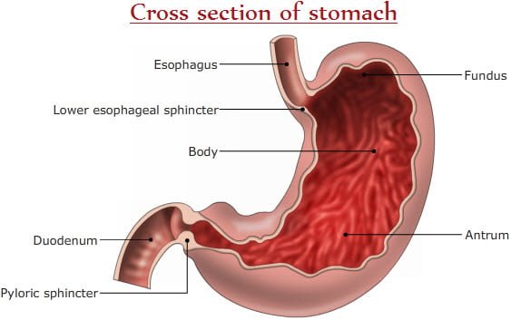
Stomach functions as a reservoir of food. Actively involved in grinding the food to reduce their size. Initiates enzymatic digestion of food materials. Controls the rate of passage of food to the small intestine for final digestion and absorption. It Produces the intrinsic factor for the absorption of vitamin B12 from the intestine, which involves in hematopoiesis.

- Based on structure and function of stomach, domestic animals fall into two general classes.
- Non-ruminants/ simple stomach animals – Horse, cat, dog, and pig.
- Ruminants – Cattle, sheep , goat, camel and buffalo
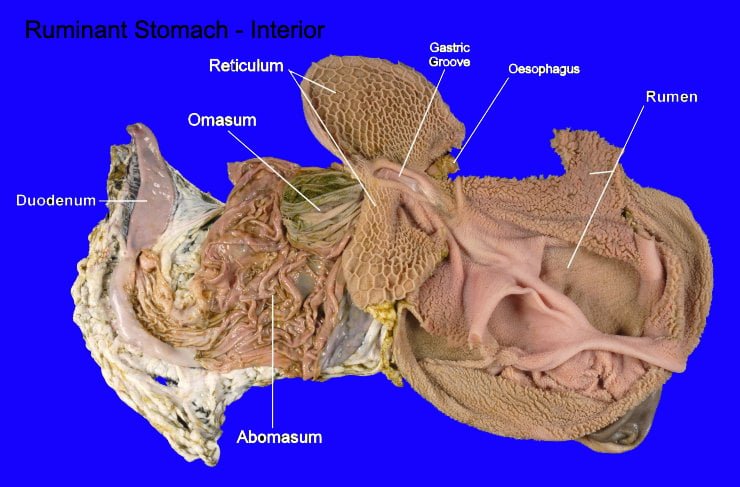
The stomach of non-ruminants is simple consisting of only one compartment, whereas the stomach of ruminants is complex, consisting of four compartments (rumen, reticulum, omasum and abomasum) of which only abomasum secretes the gastric juice. The stomach is a hollow, sac like organ made up of four layers – serous, muscular, submucosa and mucosa from outside to inside.
- The stomach mucosa of simple stomach animals, is divisible into oesophageal (glandless)region and glandularregion. The glandular area includes cardiac, fundic (parietal)and pyloric regions.
- In horse, oesophageal region is extensive up to 1/3 to 1/5 of the surface area of stomach. In the glandular region, the cardiac gland zone is very narrow, while the fundic gland zone is very wide.
- In pig, the oesophageal region of the stomach is limited to a small area around the cardia. The cardiac gland zone is very extensive, whereas the fundic and pyloric gland zones are similar to those of horse.
- In dogs, the oesophageal region is absent, the cardiac glands are found as a narrow zone scattered along the lesser curvature of the stomach around the cardia. The fundic glandular zone is extensive occupying about 2/3 of gastric mucous membrane.
Ruminant Stomach
- Ruminants are animals capable of regurgitating their food from their stomach and re-masticate them.
- Capacity of the stomach varies with age and size of the animal.
- All herbivorous animals have spacious compartments in their G.I tract. It favors retention of bulky fibrous plant for soaking, mixing and microbial fermentation.
- In ruminants and kangaroo, stomach(Rumen) provides an additional space for microbial fermentation.
- Bacteria, protozoa and fungi in the rumen are responsible for extensive fermentative digestion in the rumen. It is supported by the mechanical activity of the three compartments(rumen, reticulum and omasum).
- Only the abomasum, the true stomach secretes gastric enzymes and HCl. Ruminants are animals which can regurgitate and re-masticate.
- Abomasum is the largest compartment in new born ruminants.
- As age advances, the rumen and reticulum grow at a faster rate than abomasum.
- In adults, rumen and reticulum occupies 69%, omasum 8% and abomasum 23% of the stomach portion.
- Omasum is not well developed in sheep and goat, it is absent in Camel and Llama (Tylapoda)
- Oesophagus opens into the rumen through cardia.
- Rumen has two sacs, dorsal and ventral sacs that are freely communicating with each other.
- Rumen is connected to reticulum by rumino–reticular folds.
- Reticulum is communicated with omasum through reticulo-omasal orifice.
- Oesophageal/reticular groove extends from the cardia to reticulo–omasal orifice.
- It is a gutter like invagination. This groove is more functional in young ruminants.
- During suckling the receptors in the pharynx and mouth get stimulated causes reflex closure of reticular groove to conduct liquid and milk directly from the oesophagus into reticulo–omasal orifice, bypassing rumen and reticulum.

In calves, reticular groove/oesophageal groove acts as a bypass route for the passage of milk directly from the oesophagus into the omasum and abomasum. Closure of this groove is mediated by behavioral, psychological response and also by chemicals. Administration of chemicals like NaCl, NaHCO3, CuSO4 and sugar solutions reflexly close this groove, but CuSO4 is less effective in calf and older ruminants, but more effective in sheep. Sodium salts like NaHCO3 (60ml of 10% solution) stimulates closure in calves.
Liver
Liver and gall bladder represent accessory structures of the digestive system. Metabolic organ, participate in digestion, metabolism and also in circulatory/immune system. Special and unique organ as it receives double blood supply, seat of metabolism and is of diagnostic purpose.
Pancreas
Representing one of the accessory structures of the digestive system secretes clear, colorless liquid representing water, enzymes and salts like sodium bicorbonate.
Small Intestine
Small Intestine has 3 portions Duodenum, Jejunum and Ileum. Concerned with digestion and absorption.
Large intestine
Small intestine has 3 major portions viz. cecum, colon and rectum. Main function includes formation of certain vitamins, completion of absorption, formation of dung and its expulsion.
Gastro-intestinal motility
The walls of GI tract are muscular and capable of generating slow waves of electrical depolarization which are originated by the presence of food/ingestion as a stimuli from various areas of gut lumen. These motility aims to propel ingest from one area to the other, to promote digestion and to aid absorption. Movement popularly known as motility are of propulsive, mixing.