TABLE OF CONTENTS
Bone Development and Ossification: Intramembranous vs Endochondral Processes
Bone development is the process by which bones form, grow, and mature from embryonic mesenchyme, ensuring the formation of a functional skeletal system. A key part of this process is ossification, also known as osteogenesis, which involves the transformation of mesenchymal cells into bone-forming cells (osteoblasts) that produce and mineralize the bone matrix.
Ossification
Ossification is the process of development of bone from the mesenchyme. In the embryonic life, the future elements of the skeleton are derived from the mesenchyme, which is derived from the mesoderm.
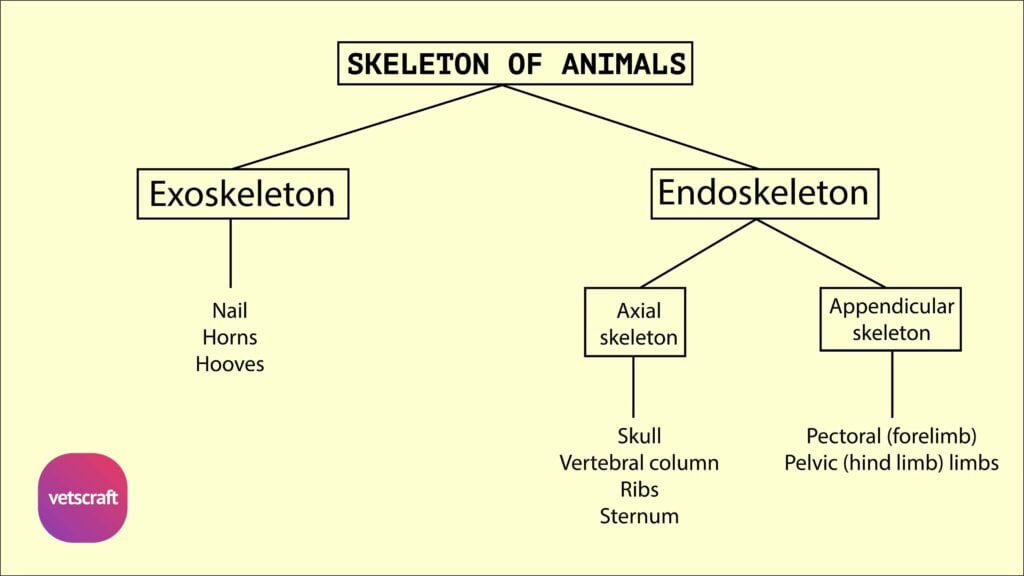
The development of bone during the fetal stage of development occurs by two processes-
- Intramembranous ossification
- Endochondral ossification
1. Intramembranous Ossification
The flat bones of the skull are developed by ossification in a membranous layer formed by the condensation of mesenchymal tissue. Hence, this process is being termed as intramembranous method of ossification. The bones developed by this method are termed as membranous bones.
The steps in intramembranous ossification are-
- Development of ossification centre
- Calcification
- Formation of trabeculae
- Development of periosteum
At a point (centres of ossification), osteoblasts are differentiated from mesenchymal cells. A meshwork of collagen fibers produced by the osteoblasts appears between the cells. It becomes vascularized by capillary network. The osteoblasts produce other organic intercellular substances like mucoprotein, glycoprotein, mucopolysaccharides etc. This organic non-calcified matrix is known as osteoid. Now the matrix is calcified by the osteoblasts. Few osteoblasts become entrapped by the surrounding matrix and are transformed into osteocytes.
Simultaneously, other osteoblasts proliferate by division and are arranged in radiating manner from the centre. Thus trabeculae are formed between the cells. The trabeculae join each other to form cancellous bone. The osteoblasts surrounding the bony spicules deposit more bones to the free ends and sides and thus calcification is spread and the bone becomes compact. The periosteum is developed from the condensation of the mesenchyme.
2. Intracartilaginous Ossification or Endochondral Method of Ossification
The bones of appendicular skeleton, vertebral column, ribs etc., are developed by intracartilagenous or endochondral method of ossification . In this method, a cartilage model of future bone is formed at first and then it is replaced by bone. Bones developed by this method are termed as cartilaginous bones.
The process of ossification does not take place simultaneously all over the cartilaginous or membranous representative of a future bone. It begins at one or more points known as centres of ossification, from which it extends until the central soft tissue is replaced by bone. In a long bone, there is a primary centre of ossification for the diaphysis and secondary centres appear for the epiphysis.
Additional centres may appear in some bones for the various processes present in the bone. The number of centres is different for the various bones in the body, but the number for each bone is constant in a particular species.
Intracartilaginous ossification or endochondral method of ossification process occurs in stages-
Stage-I
At the beginning, a cartilage model is formed by the condensation of the mesenchymal tissue. Perichondrium appears around the cartilage. The cartilage cells (chondroblasts) at the mid section of the model proliferate by mitosis and are arranged in rows towards the ends. They mature and hypertrophied. The hypertropic cells produce alkaline phosphatase and precipitate calcium salt at the matrix.
The surrounding calcification cause death of the cartilage cells and thereby form spaces-the primary areolae . This zone is known as primary ossification centre. At the same time osteoblasts appear at the inner layer of perichondrium and form subperiosteal collarbone around the primary ossification centre.
Stage-II
At this stage the collar bone is eroded by the increased activity of the subperiosteal osteoclasts and periosteal buds containing osteoblasts, osteoclasts and blood vessels enter into the primary ossification centre.
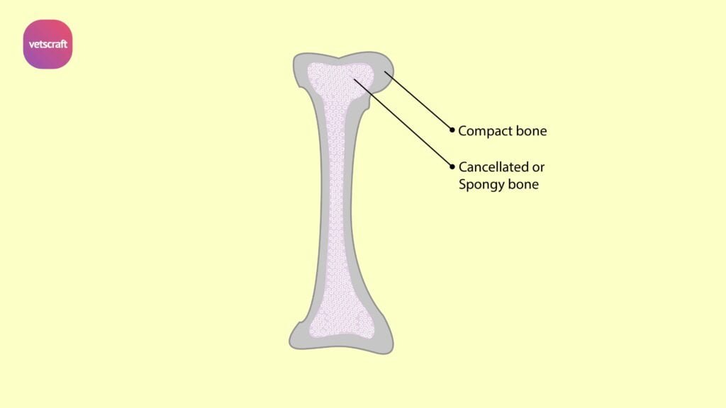
The osteoclasts absorb the irregular calcified mass and form secondary large areolae . These secondary areolae lead to the formation of marrow cavity, which subsequently becomes filled up by the bone marrow.
Stage-III
Stage-III is known as stage of true bone formation. Here the osteoblasts appear and lay down lamellated bone. Subsequently a number of longitudinal grooves proliferate and enclose a small blood vessel, which already developed along the periosteum and convert the grooves into tunnels. The lining osteoblasts of the tunnel convert the tunnel into Haversian system by proliferation and differentiation into osteocytes.
The whole process is repeated again and again and the ossification extends longitudinally. Simultaneously new bones are formed under the periosteum by appositional method.
Secondary ossification centres appears at the ends of the cartilagenous model at birth. This zone is called epiphysis. Ossification occurs in the similar way in both directions.
Growth of Bones
Flat bones of the skull increase in size by continued marginal ossification from connective tissue at the site of later sutures.
Both cartilage and membranous bones grow in thickness through further deposition of matrix at their outer surfaces.
At the ends of a long bone a layer of cartilage does not ossify and remains as articular cartilage throughout life.
In growth phase, a portion of the cartilagenous model remains as epiphyseal cartilage cartilage between the epiphysis and diaphysis.
The epiphyseal cartilage helps in longitudinal growth of the bone and is replaced totally by bone when the growth is complete. Therefore the length of the bone increases with the growth of the epiphyseal cartilage. But with the advancement of the age, the growth of this metaphyseal cartilage slows down and the calcification becomes more rapid. So the whole epiphyseal cartilages become ossified and growth in length ceases.
The width increases by the deposition of subperiosteal membrane bone.
Factors Influencing the Growth and Development of Bone
Calcium phosphorous, vitamin D, vitamin C, Alkaline phosphatase, parathyroid hormone, growth hormone of pituitary, thyroid hormone and vitamin A are factors influencing the growth and development of bone.