TABLE OF CONTENTS
Conduction system of heart in animals
The specialized excitatory and conduction fibres show feeble contractions only because of very few contractile fibres, but they conduct impulses very rapidly through out the heart.
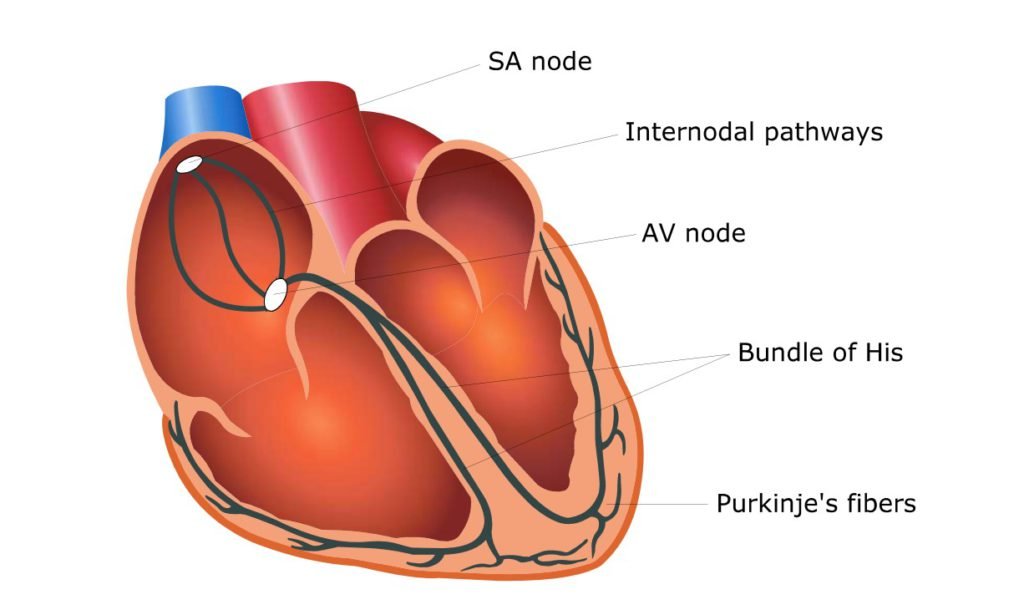
In mammals, S.A. node (Sino auricular node) is the specialized structure of the cardiac muscle located at the junction of the right atrium and cranial vena cava. It has an inherent property of generating its own action potential at periodical interval because of low resting membrane potential (- 55 mV).
The SA node normally controls the rate of the heart; hence it is called as pace maker. The pacemaker dominates the normal rate and rhythm of the heart. In sub mammalian species, frog, this function is taken up by sinus venosus. This rhythm is called as ectopic rhythm.
Most of the cardiac fibres including the conduction system have the ability of self-excitation and can produce automatic rhythmical contractions. In some pathological conditions of the mammalian heart, the excitatory impulses originate outside the S. A. node referred as ectopic foci in which the heart rate will be less than normal.
S.A. node spreads its impulse through atrial muscular wall and interatrial bundles to A.V. node (atrio -ventricular node) which lies in the septal walls of the right atrium cranio dorsal to tricuspid valve.
It conducts the action potentials to common bundle of His or AV bundle which then runs into the ventricular septum where it divides into a right and left bundle branches that run underneath the septal endocardium. At the ventricular apex, these branches finally terminate as purkinje fibres form a net work of conductive system in the ventricular muscle.
The AV node and the AV bundle is the only route for the conduction of impulse from atria to ventricles. In the conduction system, the AV node shows a delay in the propagation of the action potential, the nodal delay for a period of 50 – 150 ms.

Velocity of conduction
| Atrial muscles | 0.05 – 0.1 m/sec |
| A.V. node | 0.02 m/sec |
| Bundle of His and Purkinje fibres | 1.5-4.0 m/sec |
| Cardiac muscles | 0.3-0.5 m/sec |
The right atrium begins to depolarize about 0.01 sec before the left atrium. Conduction velocity is fastest in Purkinje fibres but slowest in A.V. nodal fibres.
The A.V. node delays allow the atria to discharge their blood into the ventricles before ventricular systole. Of the two syncytia of the heart, the atrial syncytia cause the atrial contractions a short time ahead of the ventricular contraction.