Axial Skeleton
Veterinary AnatomyThe axial skeleton of domestic animals consists of the skull, vertebral column, ribs, and sternum.
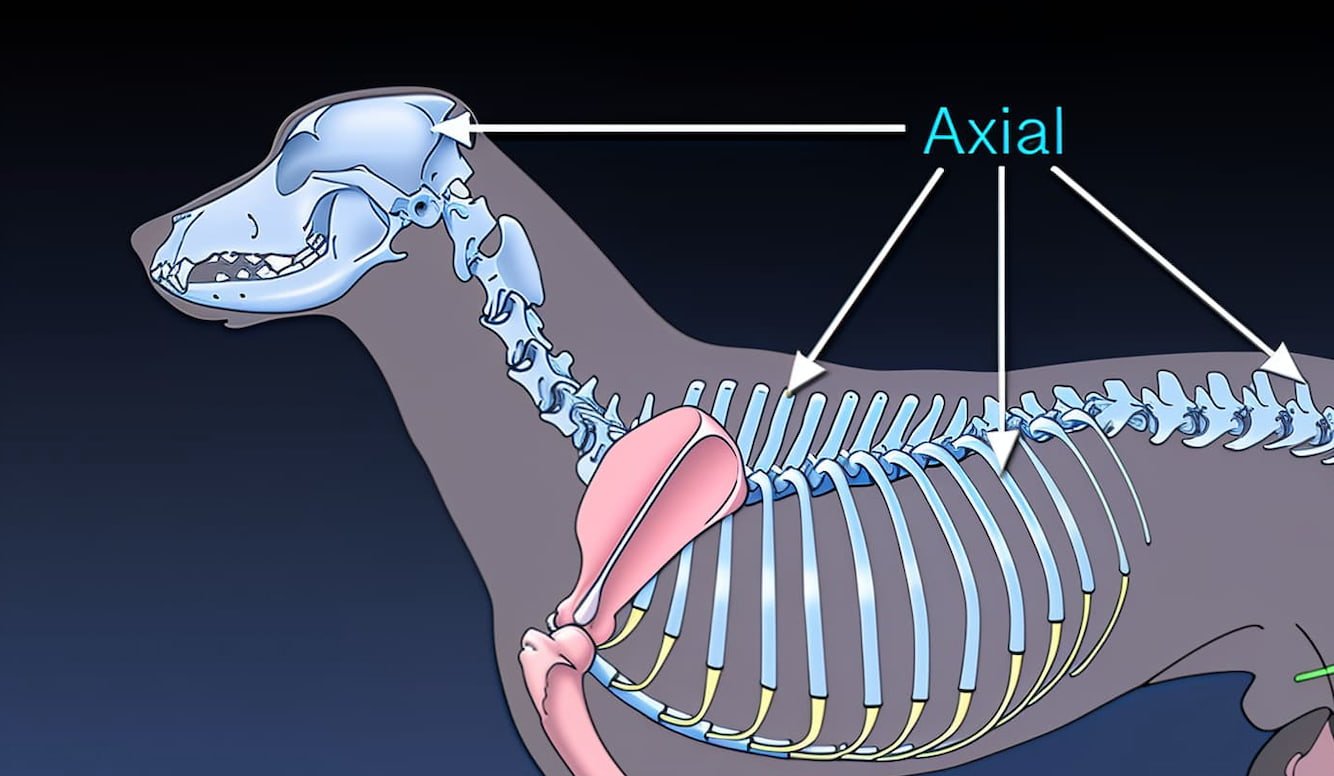
The axial skeleton of domestic animals consists of the skull, vertebral column, ribs, and sternum.

The digits of the hindlimb in animals are the terminal segments of the pelvic limb, commonly referred to as the toes. These digits are made up of phalanges (bones of the toe) and often end with claws or hooves, depending on the species.

Metatarsal bones are the long bones located in the hind limb between the tarsal bones and the phalanges (digits). They form the skeleton of the metatarsus, which corresponds to the foot region.

Tarsal bones are a group of small, irregular bones that form the hock joint or ankle region in animals. These bones connect the lower leg (tibia and fibula) to the metatarsal bones of the foot and play a crucial role in supporting body weight, providing flexibility, and enabling movement of the hindlimb.
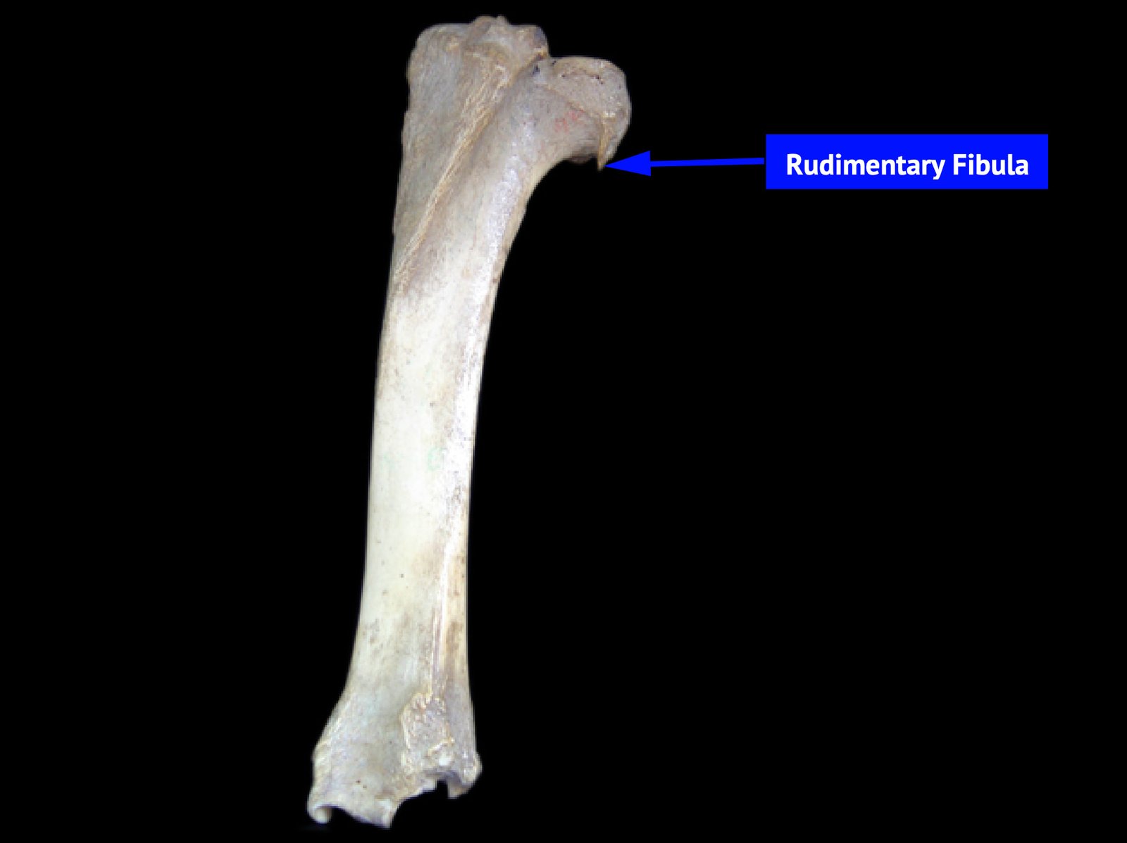
The fibula is a long, slender bone located on the lateral side of the hindlimb, running parallel to the tibia. In most animals, it plays a secondary role in weight-bearing but contributes significantly to the stability of the hock joint.

The tibia is a long, weight-bearing bone of the hindlimb in vertebrate animals. Commonly known as the shin bone, it lies between the stifle joint and the hock joint and plays a crucial role in locomotion and support.

The patella bone, also known as the kneecap, is a prominent sesamoid bone that develops within the tendon of the quadriceps femoris muscle.

The femur is the longest and strongest bone in the body, forming the skeleton of the thigh region in animals. It extends from the hip joint (acetabulum) to the stifle joint (knee), connecting the pelvic limb to the lower leg.
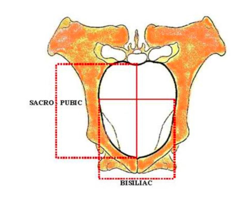
The bony pelvis of domestic animals consists of the sacrum, the first three coccygeal vertebrae, and two os coxae formed by the ilium, ischium, and pubis.

The os coxae is commonly known as the hip bone. It is a large, flat, and irregularly shaped bone that forms the lateral and ventral walls of the pelvic cavity in vertebrates.