TABLE OF CONTENTS
Anatomy of Tibia Bone in Animals: Ox, Horse, Sheep, Goat, Pig, Dog, Rabbit, and Fowl
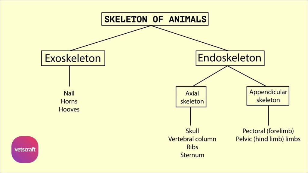
The tibia is a long, weight-bearing bone of the hindlimb in vertebrate animals. Commonly known as the shin bone, it lies between the stifle joint and the hock joint and plays a crucial role in locomotion and support.
In birds like fowl, the tibia fuses with proximal tarsal bones to form a specialized structure called the tibiotarsus.

Tibia Bone of Ox
The tibia bone of the ox is a long bone placed obliquely downward and backward between the stifle and hock joints. It consists of a shaft and two extremities.
Shaft
The shaft is three-sided above and becomes smaller and flattened below. It has three surfaces and three borders.
The lateral surface is wide above and inclines gradually to the front of the bone distally. It is covered by the tibialis anterior.
The medial face is subcutaneous, broad above, and is slightly convex and rough for the medial ligament of the stifle, sartorius, gracilis, and semimembranosus.
The posterior face is flat. The upper fourth of this surface has a triangular area marked by the popliteal line for the popliteus. The rest of this surface is marked by rough lines for the deep flexor.
The anterior border is prominent in its upper third, forming the tibial crest. It presents, on its medial aspect, a rough prominence for the semitendinosus. The rest of its extent is rounded and indistinct.
The lateral border is concave and has the fibrous part of the fibula applied against it in life. The medial border is thick and rounded in its upper fourth for the popliteus.
Proximal Extremity
The proximal extremity is large and is made up of two condyles, a tuberosity, and a spine.
The tuberosity is anterior; it is continuous distally with the tibial crest and serves for the attachment of the straight ligaments of the patella. Between the tuberosity and the lateral condyle is the sulcus muscularis for the passage of the tendon of origin of the complex muscle.
The condyles are medial and lateral. Each presents a saddle-shaped articular surface above for the corresponding condyle of the femur and the meniscus. The condyles are separated behind by the popliteal notch, on the medial aspect of which is a tubercle for the posterior cruciate ligament. The lateral condyle has the rudimentary fibula fused with it on its lateral aspect and serves for the attachment of the lateral ligament of the stifle.
The tibial spine is placed between the condyles, whose articular surfaces are continued onto the spine. It is bifid at the summit. In front of and behind the spine are depressions for ligaments.
Distal Extremity
The distal extremity is smaller than the proximal. The articular surface presents two deep sagittal grooves.
The malleoli are bony prominences on the outer margins of the sagittal grooves. The medial is smaller and fused with the distal extremity of the tibia. The medial groove is bounded on the medial aspect by the medial malleolus (which is fused to the tibia). The latter is rough medially for ligamentous attachments and articulates laterally. Its anterior part is prolonged downward to end in a blunt point.
The lateral groove is separated by a sharp ridge from an outer area, which is for the lateral malleolus. The latter completes the lateral groove.
Comparative Anatomy of Tibia Bone
The comparative anatomy of the tibia bone focuses on the structural differences and similarities of the tibia in various domestic animals, taking the ox as the reference.
Sheep and Goat
- The tibia bone of sheep and goats resembles that of the ox.
Horse
- The medial border presents, at its upper part, a small tubercle for the popliteus in the tibia of the horse.
- The popliteal line is prominent.
- The anterior tuberosity is grooved vertically.
- Below the lateral margin of the lateral condyle is a facet for the fibula.
- The grooves on the distal extremity are oblique.
- The lateral malleolus is fused to the tibia.
Pig
- The shaft is slightly curved in the tibia of the pig.
- The tibial tuberosity is grooved in front, and a narrow sulcus separates it from the lateral condyle.
- The proximal part of the tibial crest is very prominent.
Dog
- The shaft forms a double curve; the proximal part is convex medially, and the distal part is convex laterally in the tibia of the dog.
- The tibial crest is prominent.
- The facet for the fibula is on the postero-lateral aspect of the lateral condyle.
- The distal extremity presents, laterally, a facet for the fibula.
Rabbit
- The tibio-fibula is formed by the fusion of the tibia and fibula in the rabbit.
- The tibia is larger, and the fibula is thin and rod-like, situated along the lateral side of the tibia.
- The fibula is not fused with the tibia in its proximal third. Hence, the tibia alone forms the proximal articular surface.
- Distally, both the tibia and fibula share the articulation.
- The medial and lateral malleoli are in the form of small projections on the sides of the articular surface.
Fowl
- The tibia fuses below with the upper row of tarsal bones and is hence called the tibiotarsus in the case of the fowl.
- The tibio-tarsus is the longest bone in the body.
- The proximal extremity is large and irregular.
- The distal extremity comprises a trochlea behind and two condyles in front, representing the fused bones of the upper row of the tarsus.