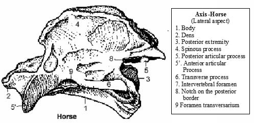
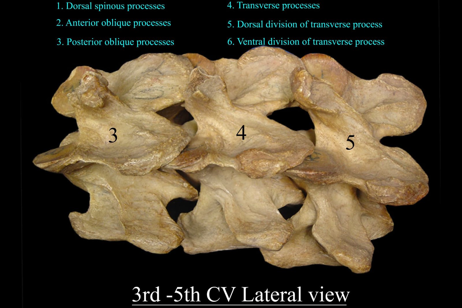
Axis of Horse - Cervical vertebrae - vetscraft

Axis

Veterinary Anatomy

Anatomy of Axis Vertebra (Vertebra Dentata) in Animals The axis vertebra, also known as vertebra dentata, is the second cervical vertebra (C2) in animals. It is uniquely adapted to support rotational movement of the head by forming the atlantoaxial joint with the atlas (C1). This joint allows the head to pivot from side to side. The axis is

Axis Read More »

Superior Posterior View of Atlas Vertebra (1st Cervical Vertebra)

Atlas

Veterinary Anatomy

The atlas is the first cervical vertebra (C1) in animals, situated immediately caudal to the skull. It articulates with the occipital condyles of the skull to form the atlanto-occipital joint, allowing dorsoventral flexion of the head.

Atlas Read More »

Tarsal bones of Domestic Animals - vetscraft - ox, horse, dog, pig, fowl

Tarsal Bones

Veterinary Anatomy

Tarsal bones are a group of small, irregular bones that form the hock joint or ankle region in animals. These bones connect the lower leg (tibia and fibula) to the metatarsal bones of the foot and play a crucial role in supporting body weight, providing flexibility, and enabling movement of the hindlimb.

Tarsal Bones Read More »

Rudimentary Fibula Bone of Ox

Fibula Bone

Veterinary Anatomy

The fibula is a long, slender bone located on the lateral side of the hindlimb, running parallel to the tibia. In most animals, it plays a secondary role in weight-bearing but contributes significantly to the stability of the hock joint.

Fibula Bone Read More »

Scroll to Top