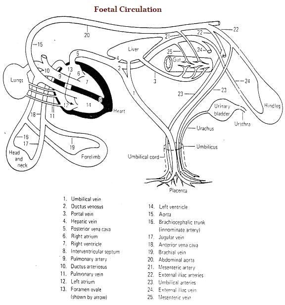
Development of Foetal circulation in animals
Oxygenated blood returning from the placenta, enters the embryo by the large umbilical vein and is conveyed to the liver. From here it is conveyed to the posterior vena cava for the most part directly through the ductus venosus and to a certain extent through the hepatic- veins.

The impure blood of the portal vein and posterior vena cava contaminates partially the large volume of Oxygenated blood from the placenta.
In sharp contrast, the blood returning from the anterior venacava is very poor in oxygen.
In the heart, the blood coming from the anterior venacava is directed into the right ventricle and through the pulmonary artery it leaves the heart.
Some of this reaches the lungs but mostly it is conveyed through the ductus arteriosus to the posterior aorta.
On the other hand the blood from the posterior venacava entering the right atrium passes mostly to the left atrium through the foramen ovale and reaches the left ventricle.
From here it is pumped into the aorta. Thus the blood reaching the heart substance through the coronary arteries and the head and neck through bicarotid trunk and its branches contains comparatively more oxygen than that which is distributed to the other parts of body through the posterior aorta.
The umbilical arteries arising from the aorta transport a large volume of this blood to the placenta for oxygenation.
At birth, the lungs become functional and placental circulation ceases. This throws some foetal vessels into disuse. The umbilical vessels pass into sudden and complete disuse.
The arteries become transformed into lateral ligaments of the bladder and the vein forms the ligamentum teres of liver.
The ductus venosus also atrophies and is transformed into the fibrous ligamentum venosus embedded in the wall of the liver.
The ductus arteriosus is transformed into the ligamentum arteriosum and the foramen ovale is obliterated and the site is marked permanently by the fossa ovalis.

