Ribs
Veterinary AnatomyRibs of domestic animals are elongated, curved bones that form the lateral walls of the thoracic (chest) cavity. They occur in pairs and correspond to the number of thoracic vertebrae.
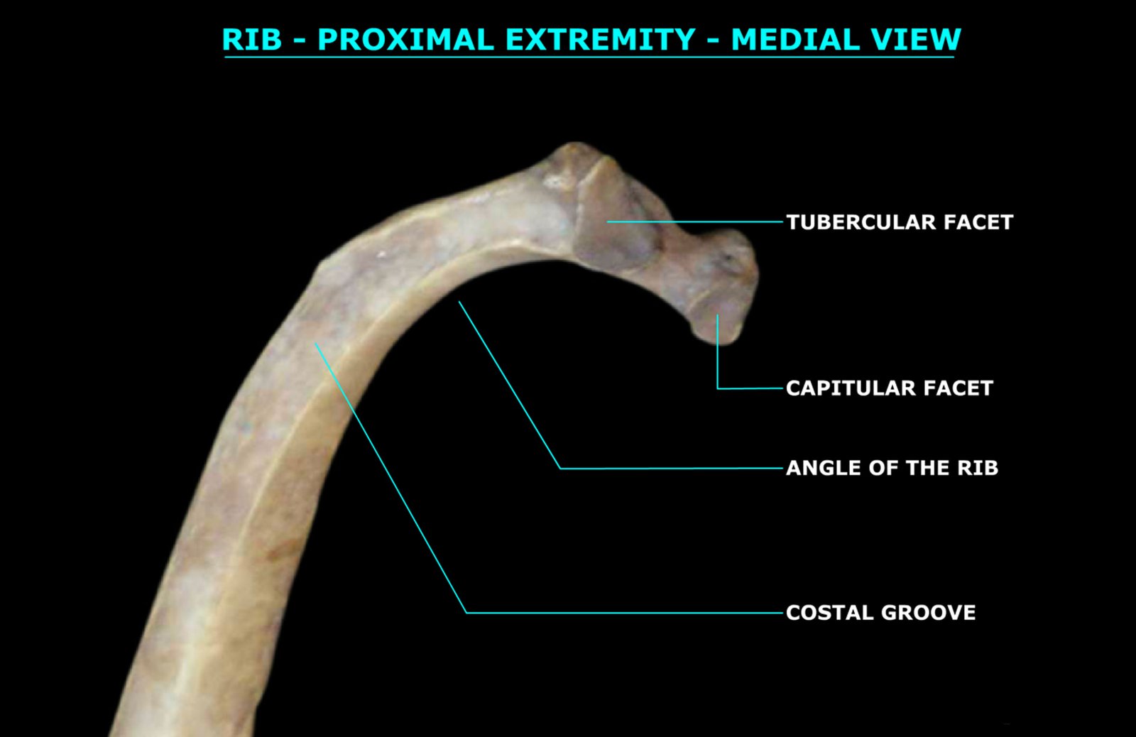
Ribs of domestic animals are elongated, curved bones that form the lateral walls of the thoracic (chest) cavity. They occur in pairs and correspond to the number of thoracic vertebrae.

The coccygeal vertebrae, also known as caudal or tail vertebrae, form the terminal portion of the vertebral column in animals. Typically, the coccygeal vertebrae become progressively smaller toward the tip of the tail, with the posterior ones often reduced to simple bony cylinders.
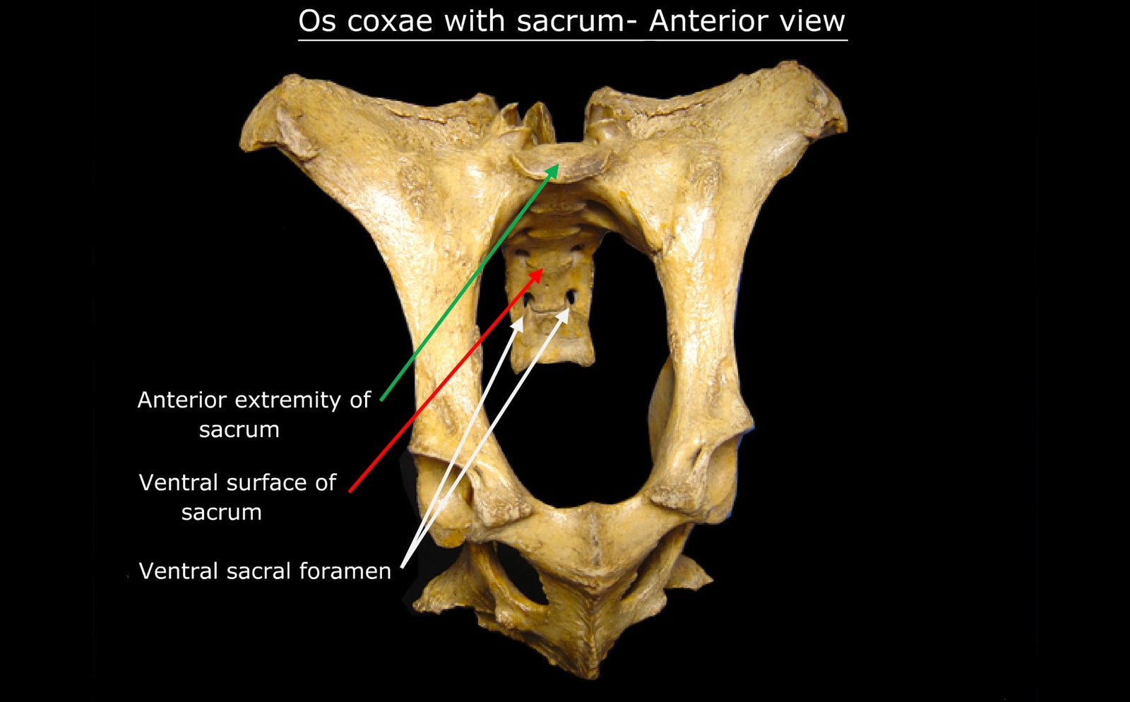
The sacrum is a part of the vertebral column in domestic animals, formed by the fusion of multiple sacral vertebrae. It serves as a strong, wedge-shaped structure that connects the spine to the pelvis and supports the hind limbs.
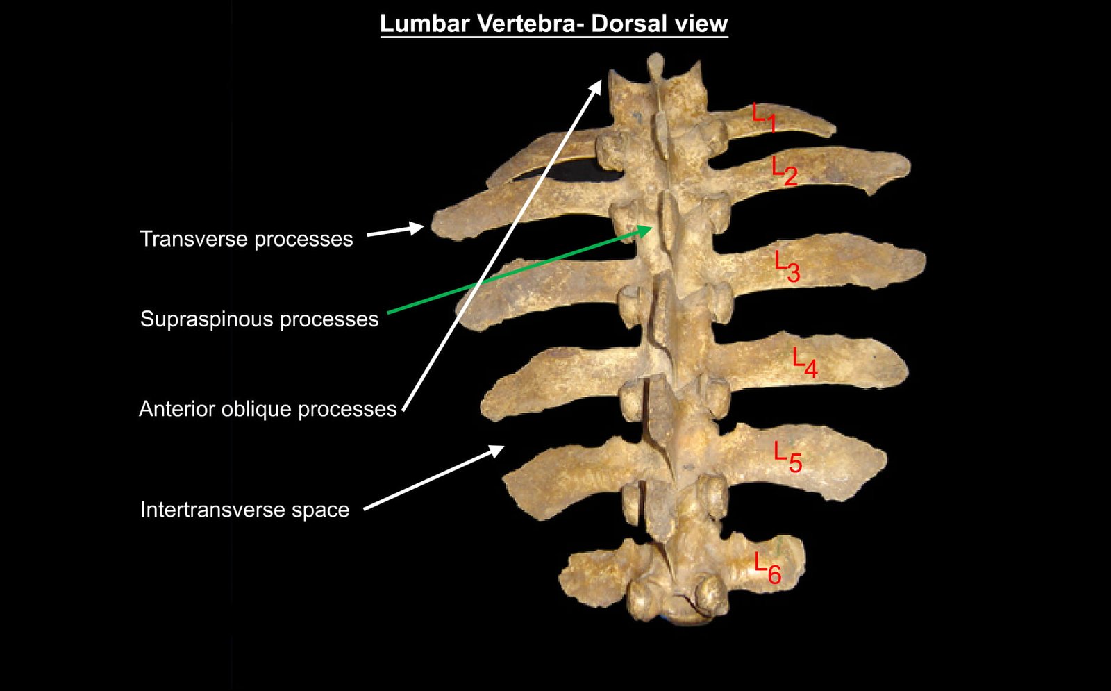
The lumbar vertebrae are the bones of the lower back, situated between the thoracic and sacral regions of the vertebral column. They are typically larger and stronger than other vertebrae, as they bear more body weight and allow a certain degree of flexibility.
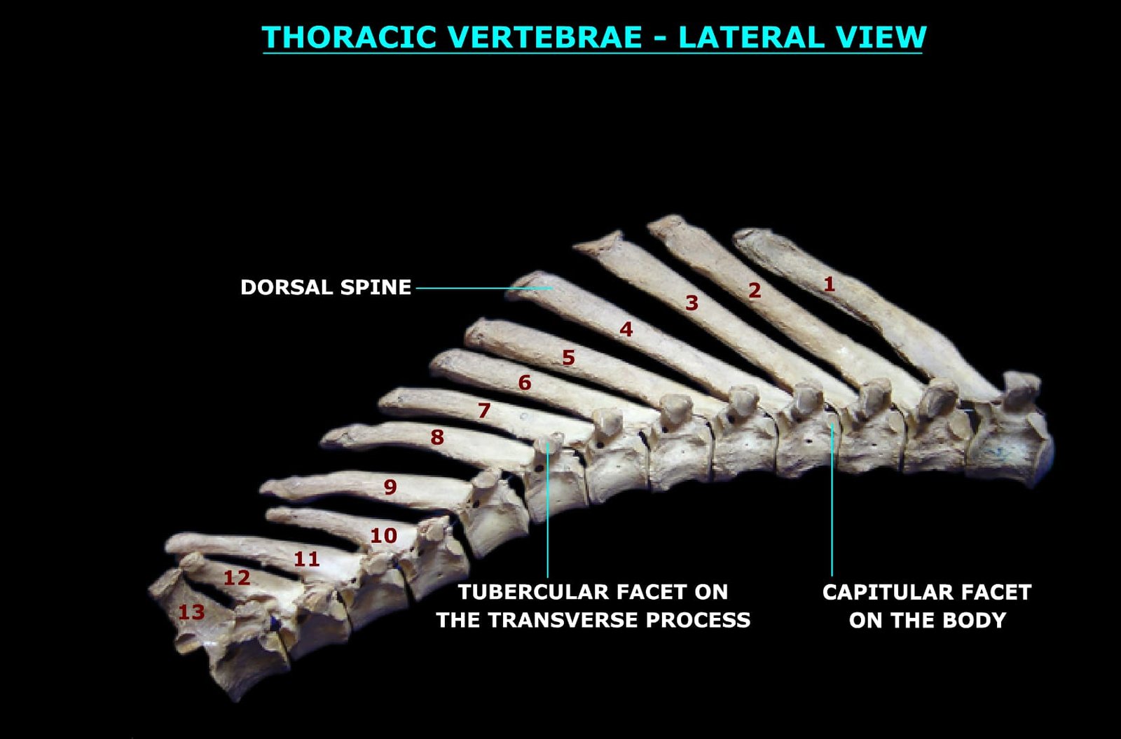
The thoracic vertebrae are the segment of the vertebral column located between the cervical and lumbar regions and are anatomically defined by their articulation with the ribs via costal facets.
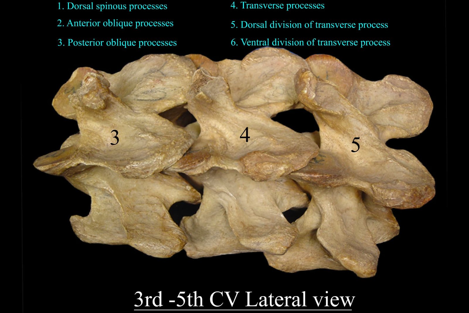
The 3rd to 7th cervical vertebrae form the caudal portion of the cervical region in animals and are crucial for supporting the neck, enabling movement, and protecting the spinal cord. These vertebrae exhibit species-specific variations in shape, articulation, and processes, which reflect the functional adaptations of different animals.
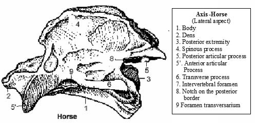
Anatomy of Axis Vertebra (Vertebra Dentata) in Animals The axis vertebra, also known as vertebra dentata, is the second cervical vertebra (C2) in animals. It is uniquely adapted to support rotational movement of the head by forming the atlantoaxial joint with the atlas (C1). This joint allows the head to pivot from side to side. The axis is

The atlas is the first cervical vertebra (C1) in animals, situated immediately caudal to the skull. It articulates with the occipital condyles of the skull to form the atlanto-occipital joint, allowing dorsoventral flexion of the head.

The cervical vertebrae form the skeleton of the neck. In the ox, horse, and dog, there are 7 cervical vertebrae. In all animals, including the fowl, the first and second cervical vertebrae, named atlas and axis respectively, are atypical vertebrae.

The vertebral column in animals is the central axis of the skeleton, consisting of a series of interconnected bones called vertebrae. It provides structural support, protects the spinal cord, and enables movement and flexibility.