Interbrain (Diencephalon)
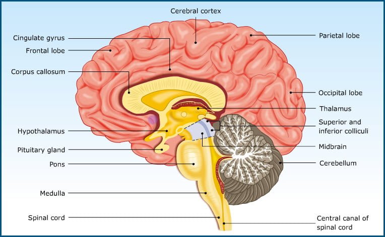
Veterinary PhysiologyInterbrain (Diencephalon) Structures including epithalamus, subthalamus (ventral area), thalamus (dorsal area), hypothalamus, pituitary gland and pineal gland constitute the Interbrain (Diencephalon). The epithalamus functions as olfactory centre. Subthalamus regulates motor activity through reticular nucleus.