TABLE OF CONTENTS
Surgical affections of teeth
Surgical affections of teeth in animals are abnormal numbers, irregularities, alveolar periostitis, dental tartar, sharp teeth, overlapping molars, mal occlusion, dental fistula, odontoma, Ameloblastoma or Adamantinoma etc.
Development of Teeth
The enamel develops from epithelium lining of the buccal cavity of the embryo.
Ameloblasts of the enamel organ form enamel for the developing tooth.
Lining the pulp cavity are specially modified connective tissue cells called odontoblasts and their function is the production of dentine.
A thin layer of bony tissue or cementum later forms on the outer surface of dentine around the root of the tooth. The dentine, cementum and pulp are derived from the mesenchyme.
Structure of Teeth
The crown is the part of the tooth projecting above the gums and the root is the part contained within the bony tooth cavity or alveolus. The crown and root meet at the neck, which is covered by the gum.
The hard portions of the tooth consist of the enamel, cementum and dentine. The dense, pearly white, outer layer of the crown is the enamel which is the hardest substance in the body. At the neck, enamel is continuous with the cementum which is a thin layer that covers the root except for the apical foramen. The cementum is bone like tissue and is difficult to distinguish from the dentine which it covers. The bulk of the tooth is formed by dentine which surrounds the pulp cavity. It is thickest in the crown and tapers to a point at the root. Its outer surface is covered by enamel in the region of crown and by the cementum in the region of the root.
The soft portion of the tooth is the pulp which is composed of sensory nerves, arteries, veins and lymphatics and primitive connective tissues which hold them together. The small apical foramen at the end of the root enables the passage of vessels and nerves in and out of the tooth.
The roots of the teeth are fairly constant. The incisor and canine teeth of both jaws have single root each. In the upper jaw, the first cheek tooth has one root, next two cheek teeth have two each and the last three cheek teeth have three roots each. In the lower jaw, the cheek teeth have two roots each, except the first and last which have one. The most important tooth clinically is the upper 4th premolar (carnassial tooth) which has two anterior roots in a transverse plane and a single large posterior root.
The outer surface of the incisor teeth is the labial surface and that of the cheek teeth, the buccal surface. The inner surface of the teeth is called as the lingual surface. The inner surface of the teeth which face the opposite dental arch is known as the occlusal or masticating surface.
The teeth are held in sockets called alveoli. The periodontal membranes serve as periosteum to the alveolar bone and provide a firm attachment between the root of the tooth and the bone. It consists of thick collagen bundles and differs from the usual periosteum in that there are no elastic fibres.
The gums (gingivae) cover the hard palate and the alveolar processes of the upper and lower jaws and surround the necks of the teeth. The gums are dense fibrous tissue and are covered with mucous membrane and are continuous with other soft tissues of the mouth.
Surgical affections of teeth
Surgical affections of teeth are–
- Developmental abnormalities of teeth
- Dentigerous cyst
- Dental tartar
- Alveolar Periostitis
- Sharp teeth
- Overlapping Molars
- Dental hook
- Wave-formed mouth
- Step-formed Mouth
- Premature wear of teeth
- Smooth mouth
- Mal Occlusion
- Shaky tooth
- Dental fistula
- Odontoma
- Ameloblastoma or Adamantinoma
- Periodontal disease
- Oral tumors
Developmental abnormalities of teeth
Developmental or congenital abnormalities of teeth are Abnormal number of teeth, Irregularities in the shedding of temporary teeth, Abnormalities of the position and direction of teeth.
Abnormal number of teeth
Supernumerary incisors and molars are frequently seen and it must be differentiated from retained deciduous teeth. There may be one or two extra teeth or a complete extra row of teeth
Due to lack of wear by not coming in contact with any apposing teeth, these extra teeth show abnormal prominences which cause injury to soft tissues are to be shortened or removed.
Irregularities in the shedding of temporary teeth
The temporary teeth may persist for a longer period. This may in turn delay the eruption of the permanent teeth or may alter their direction.
Abnormalities of the position and direction of teeth
When the teeth grow in an abnormal position or direction, they fail to come in contact with their counterparts in the opposite jaws. This causes lack of wear and the teeth become excessively long, causing injury to the soft tissues they come in contact with.
Periodical shortening of the overgrown teeth is indicated in such cases.
Dentigerous cyst
A dentigerous cyst is one containing a tooth from the bone over which it is situated. It is seen occasionally in the horse and rarely in cattle, sheep and dogs.
Dentigerous cyst develops soon after birth, along with tooth eruption and is usually noticed by about two years old. It appears initially as a soft painless swelling towards the front of the base of the ear.
Later the wall of the cyst ulcerates and then ruptures, leading to the escape of the fluid.
Passing a probe through the opening may confirm the diagnosis. As a rule, the teeth are not firmly fixed, but embedded deeply in the temporal bone. Several teeth may develop successively, following removal of a tooth.
Treatment of dentigerous cyst
Surgical excision of the lesion and try to extract the teeth without fracturing the skull.
Dental tartar
Dental tartar is a greyish brown or yellow deposit accumulating in the teeth. This condition is common in dogs and cats. Tartar consists of organic matter, bacteria, calcium carbonates and phosphates, predisposes to gingivitis and alveolar periostitis.
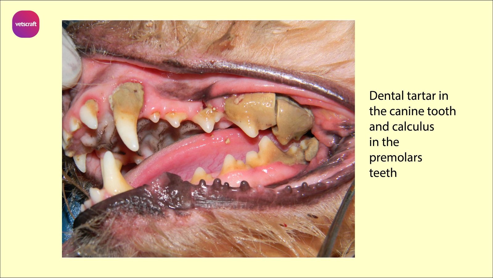
The tartar is removed with dental scalers and they are used from alveolar border to prevent injury to the gum.
In chronic cases, Dental tartar may leads to a condition called dental calculus.
Alveolar Periostitis
Inflammation of the alveolar periosteum is alveolar periostitis and it may be classified into two types- Chronic ossifying alveolar periostitis and Purulent alveolar periostitis.
The chronic ossifying alveolar periostitis is more common in horses and cattle. Suppurative or purulent type of alveolar periostitis is seen commonly in carnivores.
Chronic ossifying alveolar periostitis
Chronic ossifying alveolar periostitis is characterized by the formation of exostosis on the root of the tooth. The lower molars are more commonly affected.
The 3rd and 4th molars are more often diseased than the other teeth. The incisors are only rarely affected.
Etiology of Chronic ossifying alveolar periostitis
Inflammation of the alveolar periosteum is caused by the presence foreign body or infection.
Accumulation of food materials or tartar, fracture of the jaw involving the alveolus, caries of the tooth, excessive wear of tooth up to the level of the gum etc. exposes the alveolus to infection.
Symptoms of Chronic ossifying alveolar periostitis
Slow mastication, quidding and accumulation of food between the teeth and cheek are seen.
Food is not chewed in the affected side of the mouth. A peculiar ‘carious’ smell from the mouth is present.
Receding of the gum and change in the direction of the affected tooth as it becomes loose are also observed.
Treatment of Chronic ossifying alveolar periostitis
Treatment of Chronic ossifying alveolar periostitis is extraction of the affected tooth.
Purulent alveolar periostitis
Purulent type of inflammation of alveolar periosteum is commonly seen in dogs.
Any condition that interferes with attachment of teeth to the gums and alveolus may be considered as a predisposing factor. It is a sequel to gingivitis from any cause.
Accumulation of tartar may be considered as main cause for the condition.
Purulent alveolar periostitis condition is commonly seen in dogs maintained on soft food.
Lack of proper chewing is supposed to predispose softening of gum.
Gingivitis and alveolar periostitis in the devitalized gum tissue due to the action of micro organisms.
Symptoms of Purulent alveolar periostitis
The condition is characterized by local inflammation and pus formation.
The gum will be red, swollen and bleeds easily. There will be ulcerations on the gum and deposition of tartar on the teeth.
Slimy discharge may be seen on the gum or drooling out.
Halitosis (foul smell from the mouth) will be invariably present. Falling of the teeth will be there in due course.
Treatment of Purulent alveolar periostitis
Treatment involves scaling all the teeth and extracting the ones which are diseased, along with enough antibiotic cover.
A large number of teeth will reattach to the alveolus if the treatment is started before the condition is too advanced.
Sharp teeth
Sharp teeth are commonly seen in cattle and horses. The sharpness is seen on the outer border of the upper molars and the inner border of the lower molars.
As the upper jaw is wider than the lower jaw, the outer border of the upper molars and the inner border of the lower molars extend beyond the tables of the opposing teeth. But under normal conditions, there is more or less uniform wear of the tables because of the side to side movements of the jaws during mastication.
When the side to side movement of the jaws is restricted due to some reason, as in the case of weakness of the masseter muscles, painful lesions in the mouth, etc, the wear at the above mentioned borders is restricted. This result in extra sharp borders.
The sharp borders cause injury to the cheek and tongue and also make lateral movements of the jaws difficult.
The restricted jaw movements so caused further diminishes the wear at the already prominent borders and aggravates the condition.
Symptoms of sharp teeth
As the sharp borders of the upper molars rub on the cheek and those of the lower molars cause injury to the tongue during mastication, resulting in pain. There will be imperfect grinding of food.
The animal may hold the head to one side during chewing. Partially chewed food materials mixed with saliva may drop out of the mouth while chewing, i.e. quidding, will be invariably present.
Foaming saliva may be seen at the borders of the mouth while chewing. If the mouth is opened and examined, food materials accumulating between the cheek and molars may be seen.
The sharp edges of the teeth can be either palpated from outside or they can be detected during open mouth examination. There may be wounds or ulcers on the tongue and inner aspect of the cheeks. There will be a gradual loss of general condition of the animal due to improper feeding.
Treatment of sharp teeth
The mouth is kept open by means of an oral speculum or by holding the tongue pulled out through the opposite side and the sharp borders of the teeth are rasped.
Overlapping Molars
Overlapping Molars is also known as Shears mouth. In this condition, the outer border of the tables of the upper cheek teeth and the inner borders of the lower cheek teeth become so prominent that they overlap like the blades of the shears.
Sometimes the borders may be so sharp as to injure the opposite gum.
Treatment of Overlapping Molars consists of periodic rasping of the sharp edges.
Dental hook
Part of the table surface of a particular tooth may project due to lack of wear. This is commonly called as a dental hook.
Dental hooks may cause injury to cheek, tongue or the opposite gum. Dental hooks are commonly seen on the first upper cheek tooth and the last lower molar in herbivores.
Dental hooks can be removed by using tooth shears or may be rasped.
Wave-formed mouth
Wave-formed mouth condition, the plane of the tables of the teeth is irregular, certain teeth being very short and their opposing counterpart in the opposite jaw too long. Usually the 4th cheek teeth are affected in this manner.
The teeth become short either due to some lack of durability of the crown or due to diseases of the alveoli.
The difficulty in mastication is caused by the opposing long tooth causing injury to soft tissues.
Treatment: To avoid difficulty in chewing, a soft diet may be prescribed. Remove the sharp points and edges of the long tooth by rasping or extract the tooth. If alveolar periostitis is present, it should be treated.
Step-formed Mouth
Step-formed Mouth is also caused by over growth of individual molars. It may also result from loss of the opposing tooth.
The irregularity in the table surface is much more than in wave-formed mouth. But the line of treatment is as in the case of wave formed mouth.
Premature wear of teeth
In some individuals, the crown of the teeth becomes worn to the level of gum at a very young age. This causes pain while chewing and also causes alveolar periostitis.
There is no treatment for this condition. The wearing of teeth may be retarded by feeding on soft diet.
Smooth mouth
Smooth mouth is caused by an excessive wear of teeth. The table surfaces of teeth appear very smooth instead of having the normal rough grinding surfaces. This interferes with proper mastication and the animal loses condition.
There is no treatment for this condition. Soft diets may be prescribed.
Mal Occlusion
When the upper jaw is much longer than the lower jaw, the upper incisors overhang the lower ones. This condition is called Parrot Mouth/Brachygnathism.
In this condition, the lower incisors are likely to cause injury to the hard palate. When the lower jaw is longer than the upper jaw, the condition is called as hypognathism / prognathism / pig mouth / sow mouth. The prognathism is accepted in certain breeds like brachycephalic breeds whereas in breeds like Dachshund and Collies, brachygnathism is common and such malocclusions may be ignored.
An aberrant tooth may project into opposing soft tissue and cause pain and irritation and in such conditions, such tooth may be extracted or their rough edges may be filed.
Shaky tooth
Shaky tooth in dogs condition is generally due to the accumulation of tartar. This condition has to be differentiated from the natural shedding process of the teeth at the appropriate age.
Treatment of Shaky tooth
If tartar is removed and subsequently the mouth and the teeth are kept clean, some cases may respond positively. Remaining cases in which response is not there, dental extraction may be advised.
Dental fistula
Dental fistula is produced by the communication of the root of a tooth with the outside.
Dental fistula well discussed in Surgical affections of sinuses.
Odontoma
Odontoma is the tumor composed of tooth tissue originating from odontoblasts. It is only very rarely met with in domestic animals.
The tumor may occur in any position on the mandible or maxilla.
When the tumor is present, extraction of the tooth will be difficult or in some cases, impossible.
In such cases, curetting or chiseling out the tumorous growth under general anaesthesia is the treatment.
Ameloblastoma or Adamantinoma
Ameloblastoma or Adamantinoma tumor is not arising from the ameloblasts, but from the odontogenic epithelial remnants.
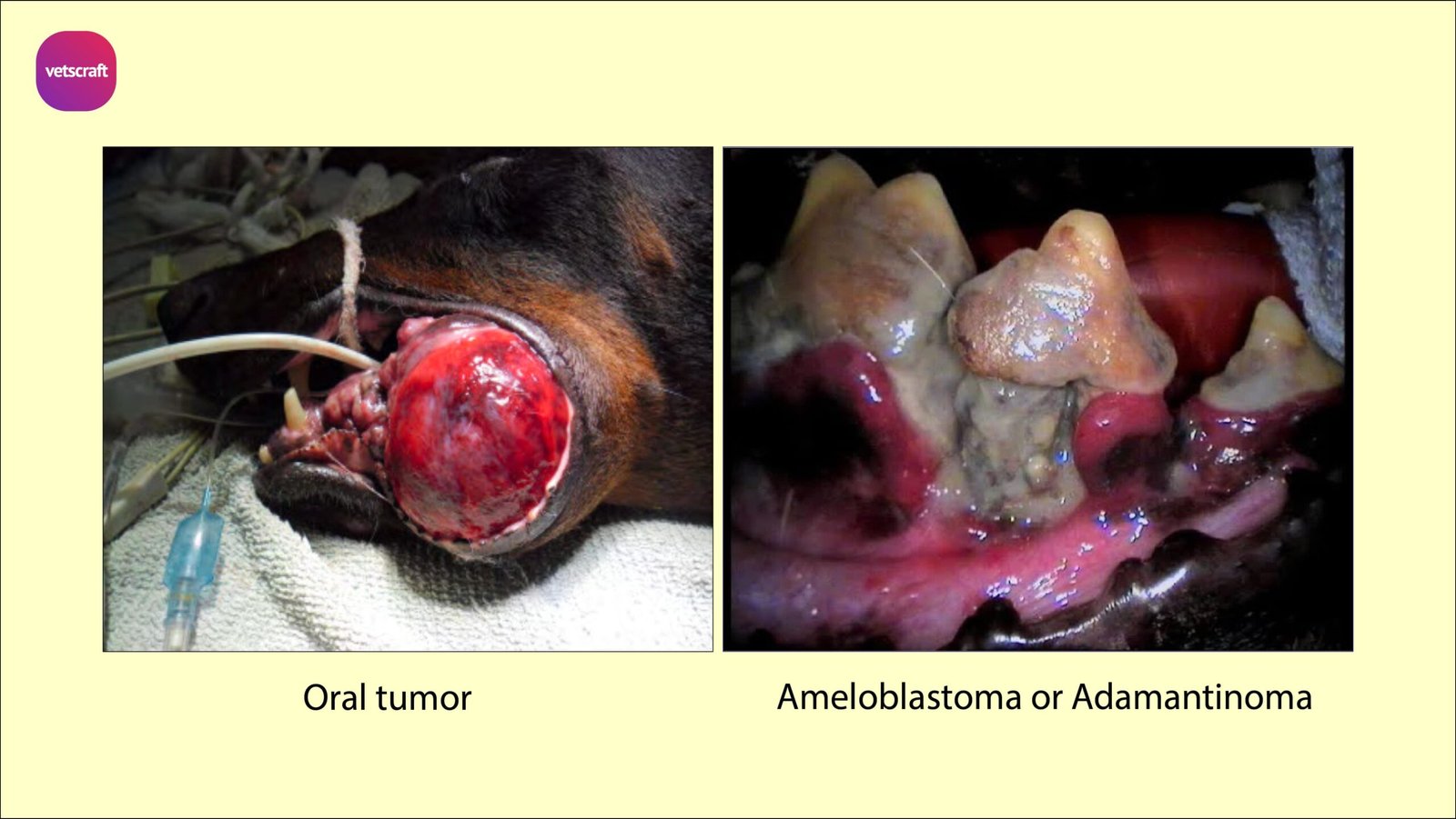
The tumor occurs sporadically in cattle, sheep and buffaloes.
Animal feels difficulty in mastication and deglutition due to abnormality and pain. In advanced cases, the incisors are displaced and embedded in the growth.
Treatment is similar to that of Odontoma.
Periodontal disease
Periodontal disease affect supporting structure of teeth like gingiva , periodontal ligament.
Supporting structures of tooth is called the periodontium. It is made up of the following parts-
- Gum (gingiva): Pale pink, firm, and does not move. It has a smooth or speckled texture. The gingival tissue between teeth is shaped like a wedge.
- Sulcus: The space between the gum and tooth
- Cementum: TheRoot surface)
- Connective tissue
- Bone : The crest of the supporting bone, which can be viewed on x-rays, is normally 2 mm below the point where the crown of the tooth meets the root (the cementoenamel junction)
Oral tumors
Oral cavity is the fourth most common site for neoplasia. The epulides or epulis (Benign neoplasm of gum and alveoli) are the most common class of oral neoplasms and accounts for 30%, less occurence is reported in cats.
They are firm gingival masses arise from the priodontal ligament.
There are three types- fibromatous,ossyfing and acanthomatous.
Oral pappilomas are benign tumours and regress spontaneously. Surgical resection is indicated only when there is dysphagia.