Hernia in animals
Hernia in animals is a condition in which part of an organ protrudes through the wall of the cavity but remains covered with skin. (If protrudes outside the skin, then it is called prolapse or evisceration).
There are various types of hernia seen in animals-
- Umbilical hernia
- Ventral hernia
- Perineal hernia
- Gut tie
- Caudal or femoral hernia
- Diaphragmatic hernia in dogs
- Diaphragmatic hernia in bovines
- Inguinal hernia
- Hiatal hernia
Umbilical hernia
Umbilical hernia is also known as Omphalocoele. this is the hernia that develops in the umbilical region. The contents usually consist of omentum and intestines. The condition is common in foals, pigs, calves and pups but rare in lambs and kids.
Umbilical hernia is comparatively more common in females than in males. The disease can be congenital or acquired. Acquired hernia is noticed few weeks after birth. Umbilical hernia may primarily be hereditary in origin due to dominant genes with low penetrance and autosomal recessive genes or due to environmental factors.
The umbilical opening in the foetus allows the passage of the urachus and umbilical blood vessels. At birth, these structures are disrupted and the opening closes around the cord.
The wound heals by cicatrisation which represents umbilicus in the later life. Acquired hernial ring may be primarily due to trauma, resection of cord too close to abdominal wall and excessive straining due to diarrhoea or constipation. Infection of the cord may also prevent natural closure of umbilicus.
Clinical Signs
- A discrete spherical swelling at umbilicus
- Hernial contents are usually fat and omentum
- Larger hernial sac contains loops of small intestine Sac is formed by skin, fibrous tissue and peritoneum
- A circular or oval hernial ring can be palpated.
- Presence of adhesions or umbilical abscess prevent reduction.
- Rarely, the contents get strangulated with symptoms of pain and intestinal obstruction
Diagnosis
- Clinical signs
- Physical examination
- Radiographs
Treatment
Umbilical hernia may be treated by various conservative or surgical methods. Conservative treatments are suitable only for a small reducible hernia.
Reducible umbilical hernia containing only a small part of the omentum or a small loop of intestine may respond favourably to abdominal pressure bandages or clamps.
- Reduce the hernia and the hernial ring to close by cicatrisation.
- Use of metal or wooden clamps: The main objective of the clamp application is to obliterate the hernial sac and to stimulate healing of the ring.
- Control the animal in dorsal recumbency
- Reduce hernial contents manually.
- Open the jaws of clamps
- Place it longitudinally and directly over the hernial ring
- Push down the hernial sac through the jaws of the clamp . Tighten the nuts of clamp to keep the clamp snugly against the abdominal wall
- The sac undergoes necrosis and sloughs down within 10-12 days.
- The skin wound heals by second intention

Hernial ring
Radical surgery
- Operation is done at the age of three months. If the swelling is too big , treatment is attempted immediately.
- Anaesthesia: General anaethesia or sedation combined with local anaethesia.
- With the animal in dorsal recumbency an elliptical incision is made on the skin over the hernial swelling.
- The incision is extended over the sac. The contents are reduced and the hernial ring is debrided and sutured.
- Preferably a synthetic non absorbable suture material is used. The ring is closed in a double breasting or overlapping pattern. Excess skin if available is trimmed before suturing.
- Hernioplasty is indicated if the hernial ring is large and weak which, could not be apposed.
Ventral hernia
Ventral or lateral adnominal hernia is the term used to describe a hernia through any part of the abdominal wall other than a natural orifice. This condition is common in horse, goat and cattle and is generally acquired in nature.
Etiology
- Any trauma– horn thrust in cattle, violent contact with blunt objects, weakening of the abdominal muscle
- Violent straining during parturition– common in sheep
- Ventral or lateral hernia is usually seen along costal arch, high or low in flank and between the last few ribs in the ventral abdominal wall near the midline
Clinical Signs
Size of the hernial ring varies in diameter. The hernial swelling is usually very prominent.
It is difficult to palpate the hernial ring in initial stages due to oedema or haematoma surrounding tissue.
Strangulation is very rare. Nature of hernial contents depends on the site of herniation.
Treatment

Herniorrhaphy
- When the hernia is harmless– herniorrhaphy is elective; not an emergency.
- It is advisable to delay the surgical repair until the inflammation subsides.
- Prolonged delay may cause complications due to adhesions between the displaced viscera and subcutaneous tissue.
- If hernia is complicated (due to incarceration or strangulation) immediate surgical intervention is required.
Surgical technique
An elliptical or linear incision is made over the hernial swelling. The contents are reduced.
The peritoneal sac is ligated or sutured close to the ring and amputated.
The hernial ring is debrided and sutured using overlapping sutures with a non absorbable suture material.

Prolene mesh
Hernioplasty using prolene mesh as onlay graft. The skin incision may be sutured with a vertical mattress sutures.

Onlay grafting
Post operative care
Amount of feed should be restricted. Supportive bandage may be placed around the abdomen. Tear of scar tissue during parturition and recurrence of hernia may occur in some cases.
Perineal hernia
Perineal hernia is most predominantly seen in old uncastrated male dogs. Though the condition is reported in bitches and cats, the incidence is rare.

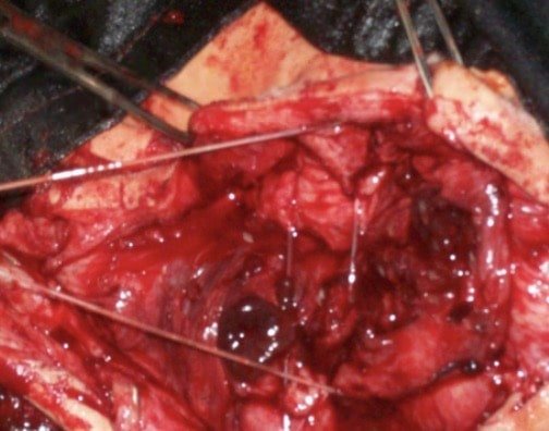
Perineal hernia in a dog
Etiology Weakening of the pelvic diaphragm and hernia can occur due to the following factors.
Hormonal disorders, Prostatic diseases, Rectal diseases and Anatomical factors – rectal deviations, diverticula etc.,
Symptoms
- Fluctuating or hard swelling ventral and lateral to the anus (in the ischiorectal fossa)
- Swelling may be unilateral or bilateral
- The hernial contents are usually rectum, enlarged prostate and perineal fat. Retroflexion of the bladder is not uncommon. Incarceration of bladder in the perineal hernia should be considered as an emergency.
- Hard swelling should be differentiated from perianal neoplasm.
Diagnosis
Diagnosis of Perineal hernia should be made from clinical signs.
Contrast radiography with barium enema will help to differentiate rectal deviation from rectal diverticulum.

Radiographic view of Perineal hernia in a dog
Ultra sound scan will be useful to identify a retroflexed bladder in animals.
Surgical management

Precise anatomical correction of perineal hernia is mandatory for successful outcome.

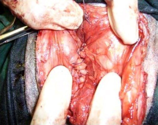
Perineal herniorrhaphy
Many techniques are available for correction of the defect-
- Conventional method
- Internal obturator flap method
Conventional method
The animal is restrained in ventral recumbency in an inclined position with the hind quarters elevated. The tail is lifted up and tied in front.
An incision is made over the swelling. The contents are reduced. The structures are identified and the correction is made in the following manner.
The medial cocygeus muscle is sutured to the external anal sphincter on the dorsal aspect. The sacro sciatic ligament is anchored to the external anal sphincter on the lateral aspect and the internal obturator muscle on the pelvic floor is sutured to the external anal sphincter on the ventral aspect.
Internal obturator flap method
Internal obturator flap method the internal obturator flap is elevated from the pelvic floor using a periosteal elevator and sutured to the medial coccygeus, levator ani and external anal sphincter.
The advantage of this technique is that it acts as a sling to suspend the rectum and does not cause undue tension on the external anal sphincter as in the conventional technique.
Recurrent perineal hernia can be corrected using techniques such as semitendinosus muscle and reinforcement with tensor fascia lata.
Gut tie
Gut tie is a type of intra abdominal hernia and it is also known as pelvic hernia and peritoneal hernia. This is formed by the passage of a portion of intestine either through a tear in the fold of serous membrane suspending the spermatic cord in the sub lumbar region or through a hernial ring like passage formed between adhesion of the cut end of the spermatic cord to the abdominal wall and the lateral abdominal wall.
Gut tie only found in bullocks; however the condition is very rare.
Etiology
Retraction of the cut end of the spermatic card into the abdominal cavity during castration and formation of adhesion between the cut end of the spermatic cord and the abdominal wall.
Gut tie occurs only on right side because rumen prevents the herniation on the left side.
Symptoms
Clinical symptoms are usually absent unless there is strangulation. When strangulation occurs animal exhibits signs of intestinal obstruction (Frequent lying down and getting up; looking towards the flank; attempting defecation)
In some cases the symptoms are noticed for a few days followed by spontaneous relief.
Diagnosis
- Pain is evinced when pressure is exerted on the right flank
- By rectal examination. The distended and herniated portion of the bowels and the stretched spermatic cord can be palpated.
Treatment
By making the animal jump from a height or making it walk down an inclination may sometime reduce the hernia.
Attempts may be made to reduce the hernia through rectal palpation.
Radical surgery is by making a right flank laparotomy and severing the adhered spermatic cord with a concealed knife.
Caudal or femoral hernia
Caudal or femoral hernia is a very rare condition and is recognized as a swelling on the muscular aspect of the thigh between sartorius and gracilis muscle.
Symtoms
Swelling in the femoral canal. The limb on the affected side is carried forward in an abducted manner during progression.
Treatment
An incision is made over the swelling and after reducing the contents the poupart or the inguinal ligament is sutured to the sartorius muscle.
Diaphragmatic hernia in dogs
Diaphragmatic hernia may be Congenital peritoneo pericardial hernia and Acquired Diaphragmatic hernia.
The signs occur at any age. sometimes will be shown while shifting to solid food. It is also noticed incidentally in thoracic radiographs or at necropsy.
Clinical signs include respiratory distress and related to the type of organ involved in herniation.
Diaphragmatic hernia in dogs is usually traumatic; like accidents or a fall from a height. The hernial content is usually a portion of omentum, stomach or liver and very rarely intestine.
The extent of herniation depends upon size and location of tear. The hernia gradually develops through a small tear due to negative pressure in the thoracic cavity and bellowing action of the abdomen during respiration.
Weakest points in the diaphragm are-
- Close to posterior vena cava
- Costal margin
- Close to the oesophagus
Symptoms
- Symptoms in congenital diapragmatic hernia, may not be noticed until the pup attains six months of age and when its starts feeding on solid foods.
- Abdominal breathing
- Peculiar cough, tendency to tire easily, unthriftiness and tucked up abdomen.
- Tendency to vomit after feeding
- Animal is reluctant to move. Remains most of the time in standing position or sitting on the haunches
- Difficulty and pain while walking down from a height
- Chronic stomach disorders
- Respiratory distress
- Gurgling sounds on auscultation of chest. Absence of respiratory sounds on affected side. More pronounced respiratory distress immediately after feeding
Diagnosis

Plain radiography in Diaphragmatic hernia in a dog

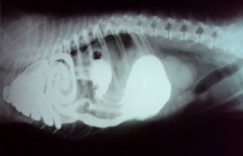
Contrast radiography showing intestines in the thorax in a dog
- History and clinical signs on auscultation – cardiac sounds are muffled
- Plain or contrast radiography
- Contrast x- ray Diaphragmatic hernia
Treatment
The condition is corrected only by surgery. IPPV The diaphragm can be approached by different approaches-
- Thoracic approach
- Abdominal approach
- Lateral Thoracic approach o Median sternotomy
- Rib splitting
The median sternotomy and rib splitting techniques are rarely used. Median sternotomy gives a very good exposure whereas the exposure to the thoracic cavity is very limited with rib splitting.
Intra operative considerations are in all the approaches the animal should be maintained under positive pressure ventilation.
Negative pressure in the thoracic cavity should be re established by aspirating the air from the thoracic cavity before the final closure.
Thoracic approach
The thoracic cavity is entered through a 6th or 7th intercostal space. The hernia is reduced and the tear in the diaphragm is sutured with a 1/0 synthetic absorbable suture material.
Care should be taken to avoid injury to lungs and other great vessels. The intercostal incision is closed including the adjacent ribs.
Abdominal approach
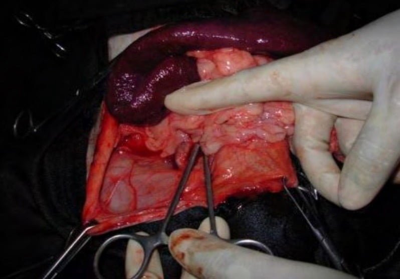
Mid line incision starting from Xiphoid backwards is made. The hernia is reduced and the tear in the diaphragm is closed in the same manner.
Diaphragmatic hernia in bovines
In cattle and buffaloes, reticulum is the common herniating organ, however the omasum, abomasum, loops of intestine, spleen or liver may also get involved.
See Diaphragmatic Hernia in Medicine by clicking here.
Etiology
Weakening of the diaphragm by lesions of traumatic reticulo-peritonitis, congenital weak points of the diaphragm and physical force like increased intra abdominal pressure during pregnancy and parturition, violent fall etc.
Clinical signs
Most affected animals develop recurrent tympany not responding to medical treatment. The tympany is mild if only a small portion of reticulam is herniated.
As more and more of the organ is herniated, signs became severe due to development of adhesions between the reticulum and other structures like lungs, pericardium, thoracic wall and hernial ring.
There will be complete or partial cessation of milk yield with passing of scanty, foul smelling pasty dung. Some cases show slight degree of melena, Regurgitation may lead to aspiration pneumonia.
Brisket edema and jugular pulse along with abduction of fore limbs may be observed. In rare cases chronic cough may be present.
On auscultation, cardiac sounds are snuffled and reticular sounds may be heard anterior the 6th rib. In untreated cases, inanition, progressive emaciation, weakness and dehydration leading to death are observed. The diagnosis is confirmed by plain and contrast radiography.
Diagnosis
- Based on clinical examination
- Left flank exploratory laparotomy may be done
- Radiography
Treatment
Treatment of Diaphragmatic hernia in bovines is only surgical correction.

The first step is to evacuate the contents of the rumen and reticulum by rumenotomy and cud transplantation. Then for animal is kept on I/v fluids for 48 hours and thereafter in the soft diets with fluids, surgery to correct hernia may be delayed 3-4 days.
The common approaches for diaphragmatic hernia are abdominal and thoracic. Irrespective of the approach, proper ruminal evacuation and assisted ventilation during herniorrhaphy are required for successful procedure.
Congenital Diaphragmatic hernia or Peritoneo-pericardial hernia
Congenital Diaphragmatic hernia or Peritoneo-pericardial hernia is a common condition due to the failure of development of septum tranversium.
Clinical signs includes vomiting, anorexia, lethargy and diarrohea and dyspnoea is infrequent.
On physical examination, heart sounds are muffled and intestinal sounds are heard in the ventral portion of the chest.
Radiography confirms the diagnosis. The hernia is repaired through ventral midline laparotomy.
Traumatic Diaphragmatic hernia
Traumatic Diaphragmatic hernia is caused usually by blunt trauma, especially automobile accidents.
The tear can occur anywhere in the diaphragm or the diaphragm may get separated from its attachment to the ribs.
The clinical signs may vary from absence of symptoms to severe dyspnoea Also, signs vary with the location and the organ terminated.
Other conditions such as rib fracture, penumothroax, lung contusion or shock also may be present.
Due to dyspnoea, the animal (dogs) rest in sitting position with the elbow abducted. On physical examination, intestinal sounds may be heard in the thorax, the heart may be displaced and the heart sounds snuffled.
Careful palpation may reveal reduction in volume of abdominal viscera or abnormal position of the organs.
Lateral and ventro-dorsal radiographs will confirm diagnosis, contrast radiography with barium meal will help in diagnosis of herniation of stomach or intestional loops.
Clinical signs are not exhibited at the time of original injury. But it may develop gradually and show clinical signs such as intermittent vomiting, anorexia, jaundice, dyspnea, reduced exercise tolerance etc.
Surgical correction is the only treatment.
Inguinal hernia
Protrusion of an abdominal organ through the inguinal canal is called inguinal hernia (Bubonocele). If the hernial contents extend into the scrotum in male animals the condition is called as scrotal hernia.

Inguinal hernia in a dog
Incidence of Inguinal hernia are common in Bitches, horses, bulls and pigs.
Inguinal canal is an oblique (slit like) canal between the abdominal muscles connecting the external and internal inguinal rings.
The canal acts as a passage for structure like spermatic cord in males and external pudic artery in females.
Internal inguinal ring is longer than external inguinal ring.
Etiology
Inguinal hernia may be congenital or acquired – accidental slipping causes streching of the hind limbs outwards which may dilate inguinal canal.
Symptoms
- In bitches appreciable swelling is noticed in the inguinal region. Difficulty in defecation.
- In large animals swelling in the inguinal canal at the neck of scrotum.
- Unilaterally enlarged scrotum; affected bulls or stallions may be reluctant to serve
- Refuse to move due to pain
- Abduction of hind limbs
- Systemic signs are evident only when the hernia gets strangulated
Inguinal hernial contents include intestine, urinary bladder, uterus in female and omentum.
Diagnosis
- Clinical signs
- Radiography or fluoroscopy (Hernial swelling close to or including the scrotum)
- Rectal palpation in large animals
Treatment
Inguinal hernia in small animals
A paramedian incision is made close to the inguinal swelling. The contents are reduced by gentle pressure. A kelotomy (extension of the hernial ring) may be performed if the hernial ring is small. The edges are debrided and closed using an absorbable suture material by overlapping pattern.
Inguinal hernia in large animals
After making the incision the hernia is reduced and purse string suture is applied around the tunica vaginalis as far high as possible. A portion of the omentum that is difficult to reduce can be amputated. Overlapping suture of the muscles and routine closure of the skin is made.
Hiatal hernia
Hiatal hernia is a form of diaphragmatic hernia in which the caudal end of the oesophagus and cardiac area of the stomach pass through the oesophageal hiatus of the diaphragm.
The associated sign is the oesophagitis. Treatment consists of reducing the hernia and reconstructing the diaphragm.
In cattle and buffaloes, reticulum is the common herniating organ, however the omasum, abomasum, loops of intestine, spleen or liver may also get involved.
Etiology
Weakening of the diaphragm by lesions of traumatic reticulo peritonitis, congenital weak points of the diaphragm and physical force like increased intra abdominal pressure during pregnancy and parturition, violent fall etc.
Clinical signs
Most affected animals develop recurrent tympany not responding to medical treatment. The tympany is mild if only a small portion of reticulum is herniated.
As more and more of the organ is herniated, signs become severe due to development of adhesions between the reticulum and other structures like lungs, pericardium, thoracic wall and hernial ring.
There will be complete or partial cessation of milk yield with passing of scanty, foul smelling pasty dung. Some cases show slight degree of melena, Regurgitation may lead to aspiration pneumonia.
Brisket edema and jugular pulse along with abduction of fore limbs may be observed. In rare cases chronic cough may be present.
On auscultation, cardiac sounds are muffled and reticular sounds may be heard anterior to the 6th rib. In untreated cases, inanition, progressive emaciation, weakness and dehydration leading to death are observed.
Diagnosis
The diagnosis is confirmed by plain and contrast radiography. Left flank exploratory laparotomy may be done.
Treatment
Treatment of Hiatal hernia in animals is only surgical correction.
The first step is to evacuate the contents of the rumen and reticulum by rumenotomy and cud transplantation. Then the animal is kept on I/v fluids for 48 hours and thereafter in the soft diets with fluids. Surgery to correct hernia may be delayed 3-4 days.
The common approaches for diaphragmatic hernia are abdominal and thoracic. Irrespective of the approach, proper ruminal evacuation and assisted ventilation during herniorrhaphy are required for successful procedure.