Neural Control of respiration
Pulmonary ventilation is regulated closely to maintain the concentrations of H+, CO2, and O2 at relatively constant levels while meeting the needs of the body under varying conditions. If either the H+ or the CO2 concentration increases or if the O2 concentration decreases, their levels will be returned to normal by increasing ventilation. Conversely, if either the H+ or CO2 concentration decreases or if the O2 concentration increases, pulmonary ventilation will be decreased. This regulatory mechanism is controlled by changes in tidal volume, frequency of respiratory cycles, or both.
Respiratory centres
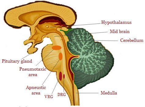
The central mediator of these changes is the respiratory center in the brain stem which has four specific regions .
The medullary rhythmicity area is located in the medulla oblongata, and it controls the basic respiration rhythm.
- It consists of two areas, the inspiratory and expiratory areas, also called the dorsal respiratory group and ventral respiratory group, respectively.
- Dorsal respiratory group: group of neurons predominately associated with inspiratory activity (lung inflation and termination of inspiration).
- Ventral respiratory group: group of neurons containing inspiratory and expiratory neurons (assist in inspiration begun by those in the dorsal respiratory group and also assists expiration).
The respiratory center is gray matter in the pons and the upper medulla, is very sensitive to the PCO2 in the arteries and to the pH level of the blood.

The CO2 can be brought back to the lungs in three different ways; dissolved in plasma, as carboxyhaemoglobin, or as carbonic acid. That particular form of acid is almost broken down immediately by carbonic anhydrase into bicarbonate and hydrogen ions. This process is then reversed in the lungs so that water and carbon dioxide are exhaled.
The medulla oblongata reacts to both CO2 and pH levels which triggers the breathing process so that more oxygen can enter the body to replace the oxygen that has been utilized.
The medulla oblongata sends neural impulses down through the spinal chord and into the diaphragm.
- There are two other ways the Medulla Oblongata can be stimulated.
- The first type is when there is an oxygen debt (lack of oxygen reaching the muscles), and this produces lactic acid which lowers the pH level. The medulla oblongata is then stimulated.
- The second type occurs when the major arteries in the body called the aortic and carotid bodies, sense a lack of oxygen within the blood and they send messages to the medulla oblongata.
The inspiratory area sends signals to the diaphragm via the phrenic nerve and to the external intercostal muscles via the intercostal nerves. These signals cause muscle contraction resulting in inspirations. When these signals cease, inspiration is concluded, which allows the diaphragm and external intercostal muscles to passively relax, during which time the elastic recoil of the lungs and thoracic walls causes the volume of the thoracic cavity to decrease.
Transection between the spinal cord and medulla oblongata stops breathing.
Although not active during quiet breathing, forceful expiration requires signals from the expiratory area that cause contraction of the internal intercostals and abdominal muscles.
Contraction of these muscles further decreases the volume of the thoracic cavity, thus increasing exhalation.
Pneumotaxic and Apneustic area
Pneumotaxic centre believed to modulate respiratory center sensitivity to inputs that activate termination of inspiration and facilitate expiration .
Apneustic centre believed to be associated with deep inspirations.
The pneumotaxic area, also called the pontine respiratory group, sends inhibitory signals to the inspiratory area. These signals primarily function to prevent overfilling of the lungs.
Conversely, the apneustic area located in the lower pons sends stimulatory signals to the inspiratory area that prolongs inspiration.
The pneumotaxic area can override the apneustic area. Impulses going to the respiratory center (afferent impulses) from several receptor sources have been identified. Importantly the Hering-Breuer reflexes.


