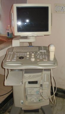
Echocardiography in animals
Echocardiography or Cardiac Ultrasonography in animals is a non invasive tool for imaging the heart of the surrounding intrathoracic structures to diagnose various cardiac diseases to assess cardiac function.

Uses of Echocardiography
- To evaluate size of cardiac chambers
- To study the thickness and movement of the wall
- To study the structure and movement of the valves
- To detect pericardial and pleural fluid
- To find out congenital cardiac anomalies
- To identify mass lesions within and adjacent to the heart
- To diagnose valvular and myocardial pathology
Principle of echocardiography
Echocardiography employs high frequency sound waves greater than 20,000 Hz. These ultrasound waves when passed through the tissues are reflected back from the tissues based on the acoustic impedance of the tissue. The amount of reflection of ultrasound waves depends on the difference in the acoustic impendance between two adjacent tissues. i.e. greater the difference, greater in the reflection. Bone tissue and air/tissue interfaces are highly reflective. Bone has a very high acoustic impedance whereas air has a very low acoustic impedence, as well as the soft tissue.
Read > Parts and mechanism of ultrasonography machine
Read > Principles of ultrasonography and its applications
Echocardiography is usually performed in the inter spaces within the cardiac windows. Ultrasound waves are transmitted through the tissue at a known speed depending upon the type of tissue through which it travels. Ultrasound obeys the laws of geometric optics with regard to reflection, transmission and refraction.
When an ultrasound wave meets an interface of differing acoustic impedance, the wave is reflected, refracted and absorbed. The intensity of the ultrasound beam decreases as it travels away from the transducer because of beam divergence, absorption, scatter, and reflection of wave energy at tissue interfaces. The largest ultrasound reflection occurs when the ultrasound beam is perpendicular to the imaged structure, creating a strong reflection or echo.
These reflected ultrasound waves are then received by the transducer and processed by the ultrasound machine to create an image. The transducer acts as a receiver over 99% of the time. The echocardiographic images obtained are displayed on the monitor and can be recorded on videotape, thermal paper, radiographic film or computer disc.
The frequency of the ultrasound waves emitted by the transducer markedly influence the quality of the image obtained and the depth of tissue that can be imaged successfully. Higher frequency ultrasound waves have a shorter wavelength and yield better resolution of small structures close to the skin surface.
However, more energy is absorbed and scattered with high frequency ultrasound and thus, high frequency transducers have less penetrating ability. Conversely, a lower frequency transducer will have a greater depth of penetration but poor resolution. The transducer selected for echocardiography should be the highest frequency available that will penetrate to the depths needed to image the heart in its entirety. Frequencies generally used for veterinary echocardiography range from 2.25-3.5 MHz for adult horses and cattle and 3.5-10.0 MHz for small animals, small ruminants, foals, calves and exotics.
Types of echocardiography
There are three types of echocardiography, used clinically- M-mode, Two-dimensional (2-D, B-mode or real time), and Doppler echocardiography.
1. M-mode Echocardiography
It is a one-dimensional (“ice-pick”) view of the cardiac structures moving over time. The echoes from various tissue interfaces along the axis of the beam are moving during the cardiac cycle and are swept across time, providing the dimension in time. The lines on the recordings correspond to the position of the imaged structures in relation to the transducer and other cardiac structures at any instance in time. More accurate placement of the M-mode cursor within the heart is performed by using the two-dimensional (2-D) real-time image as a guide. The M-mode echocardiogram uses a high sampling rate and can yield cleaner images of cardiac borders, allowing the echocardiographer to obtain more accurate measurements of cardiac dimensions and more critically evaluate cardiac motion.
Standard M-mode views are obtained from the right parasternal position. The standard M-mode views utilized in the veterinary medicine include the left ventricle (at the level of the chordae tendineae), the mitral valve and the aortic root (aorta/left artrial appendage) view.
2. Two-Dimensional Echocardiography
Two-dimensional echocardiography allows a plane of tissue (both depth and width) to be imaged in real time. Thus, the anatomic relationships between various structures are easier to appreciate than with M-mode echocardiographic images. An infinite number of imaging planes through the heart are possible, however, standard views are used to evaluate the intra and extracardiac structures. The standard views are obtained from either the right parasternal window in all species and from the left parasternal window in adult large animals or in other species when imaging the heart from the left side is desirable.
3. Doppler echocardiography
Doppler imaging allows evaluation of blood flow patterns, direction, and velocity; thus, it permits documentation and quantification of valvular insufficiency or stenosis and cardiac shunts. Estimation of blood flow and cardiac output can also be made. Doppler echocardiography is based on detection of frequency changes (the Doppler shift) occurring as ultrasound waves reflect off individual blood cells moving either away from or toward the transducer. Calculation of blood flow velocity is possible when the flow is parallel to the angle of the ultrasound beam.
Two types of Doppler echocardiography are used clinically- Pulsed wave and Continous wave.
Pulsed wave Doppler echocardiography
Pulsed wave (PW) Doppler uses short bursts of ultrasound transmitted to a point (designated the “sample volume”) distant from the transducer. The advantage of this type of Doppler is that blood flow velocity, direction and spectral characteristics from a specified point in the heart or blood vessel can be calculated. The main disadvantage is that the limited.
Continuous wave Doppler echocardiography
Continuous wave (CW) Doppler uses dual crystals so that ultrasound waves can be simultaneously and continuously sent and received. There is no maximum measurable velocity (Nyquist limit) with CW so high velocity flows can be measured. The disadvantage with CW Doppler is that sampling of blood flow velocity and direction occurs all along the ultrasound beam, not in a specified area.
Color flow Doppler echocardiography is a form of PW Doppler ultrasonography which combines the M-mode and 2-D modalities with blood flow imaging. With color flow Doppler, multiple sample volumes are analyzed along multiple scan lines. The mean frequency shift obtained from these many sample volumes is color-coded for direction and velocity. Several types of mapping are usually available. Most systems code blood flow toward the transducer as red and flow away as blue. Differences in relative velocity of flow can be accentuated, and the presence of multiple velocities and directions of flow (turbulence) can be indicated by different maps which utilize variations in brightness and color.


