TABLE OF CONTENTS
Parts and Mechanism of Ultrasonography Machine
Parts of ultrasonography machine for animals are a transducer, the processing unit, controls and the display or monitor. Mechanism of ultrasonography machine is when the transducer probe of ultrasonography machine is placed in close contact with the body surface through a coupling medium, it undergoes continuous modification which occurs through three processes-
- Absorption
- Reflection
- Scattering
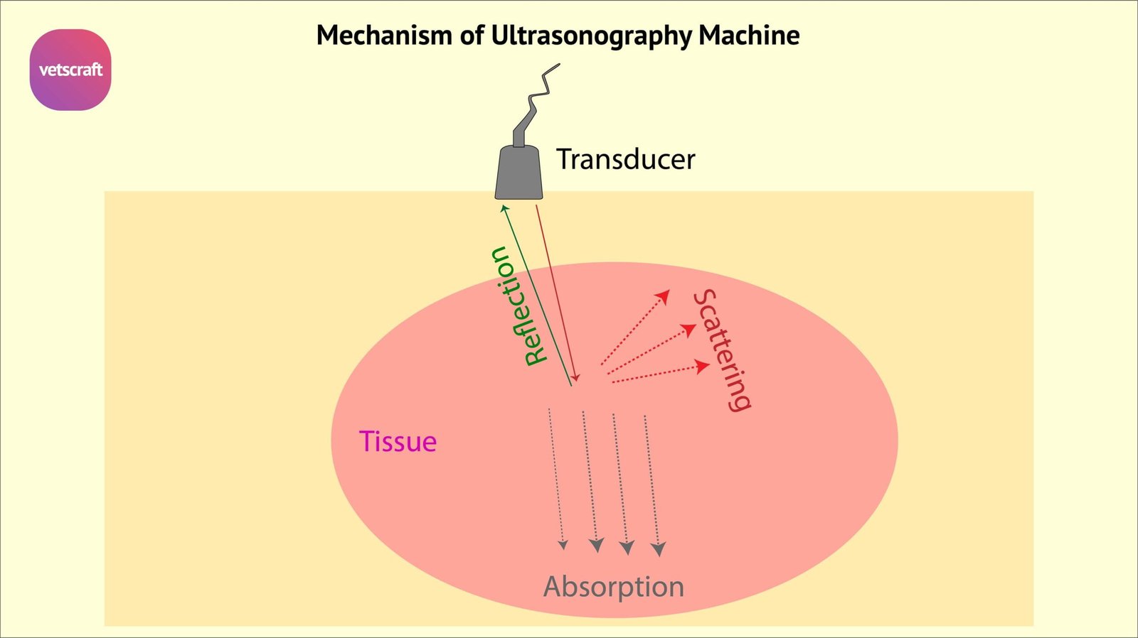
Absorption
Absorption occurs when the energy in the sound beam is absorbed by the tissues thereby converting it in to heat. It is basis for the therapeutic ultrasound.
Reflection
Reflection is the redirection of the portion of the ultrasound beam back towards the source. The reflection gives rise to different echoes. These echoes are converted by peizo electric effect in to electrical signals and displayed in to oscilloscope screen.
Scattering
Scattering occurs when the beam encounters an interface that is irregular and smaller than the sound beam. The portion of beam that interacts with this interface is scattered in all directions.
There are two other closely related phenomena, refraction and refraction of which refraction is common cause of artifacts. Larger echoes leave very little of the beam to produce echoes from another interface deeper in the tissues. This is the reason that bowel gas or bone does not allow the scanning of tissues deeper to these.
Echo Quantification (EQ)
Once the echoes are converted into electrical signals, these are processed and transformed into a visual display of measure of the amplitude of the echo. This is called echo quantification. For displaying this echo amplitude information three modes A, B and M mode are used-
- A mode (Amplitude)
- M mode (Motion)
- B mode (Brightness)
A mode (Amplitude)
A mode (Amplitude) is the simplest form of display. It displays two parameters of the echoes in the form of spike. The distance from the transducer and the amplitude or reflected strength and is rarely used.
M mode (Motion)
M mode (Motion) is similar to ‘A’ Mode but can record the motion and position of echo, used in heart valve and cardiac valve movement.
B mode (Brightness)
B mode (Brightness) is most commonly used display mode, yields two dimensional picture composed of lines per grey dots. The position of the dot is directly proportional to the actual distance the sound wave traveled through tissue. The brightness of the dot is determined by the amount of ultrasound pulse that is reflected. A cross sectional view covered by the transducer is displayed.
Transducers of Ultrasonography
The frequency of the transducer probes is determined by the times the crystals expand and contracts in one second. Transducers are in linear array, convex and sector-
- Linear array transducer
- Convex array transducer
- Sector type transducer
Linear array transducer
Linear array is a thin rectangular clip lined up side by side producing sound waves. Beam is rectangular in shape and it permits good visualisation of shape of the structure.
Convex array transducer
In a Convex array transducer, crystals are placed in curvilinear fashion. Imaging of greater area can be effected.
Sector type transducer
Sector type transducer contains single crystal which oscillates or rotates to produce a fan shaped beam. The small size gives it to access more of the thoracic and abdominal organs through a small contact area.
Real time Ultrasound
When the images displayed in B mode scan are formed rapidly and presented in sequence, the movement of organs will be viewed in real time. In order to form the sequential images, it is necessary to sweep the ultrasound beam over the tissues by either mechanical or electronic means.
The sound beam can be swept either in a arc or in a linear fashion, when it is in a form of arc, it is called sector scan.